1ZS3 image
Entry Detail
PDB ID:
1ZS3
Title:
The crystal structure of the Lactococcus lactis MG1363 DpsB protein
Biological Source:
Source Organism:
PDB Version:
Deposition Date:
2005-05-23
Release Date:
2005-08-30
Method Details:
Experimental Method:
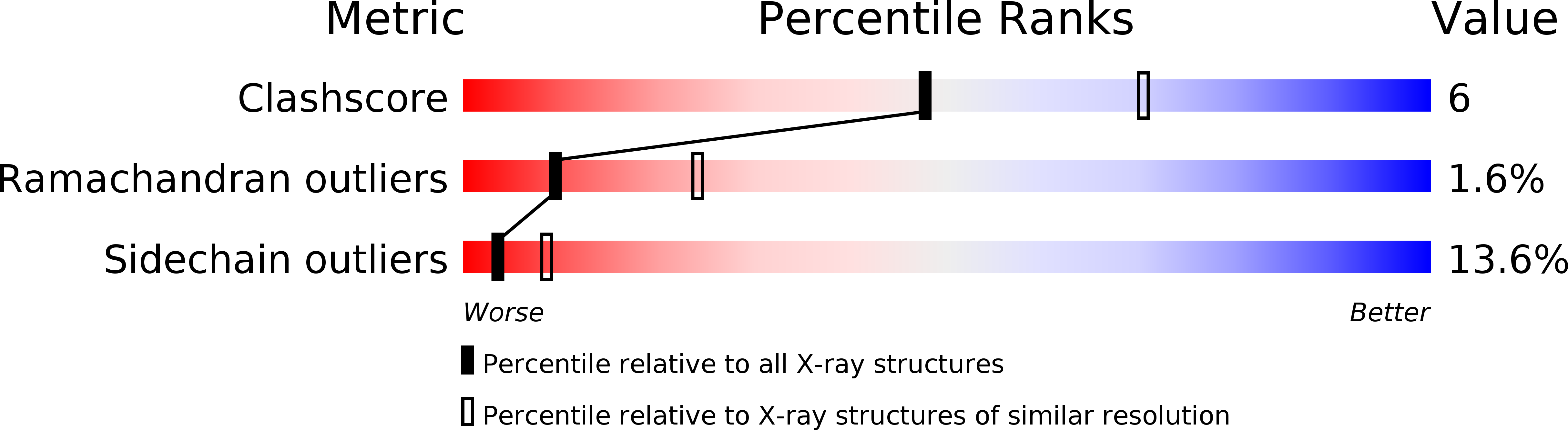
Resolution:
2.70 Å
R-Value Free:
0.25
R-Value Work:
0.20
R-Value Observed:
0.20
Space Group:
P 21 21 21
Macromolecular Entities
Polymer Type:polypeptide(L)
Description:Lactococcus lactis MG1363 DpsA
Chain IDs:A, B, C, D, E, F, G, H, I, J, K, L
Chain Length:182
Number of Molecules:12
Biological Source:Lactococcus lactis
Primary Citation
The crystal structures of Lactococcus lactis MG1363 Dps proteins reveal the presence of an N-terminal helix that is required for DNA binding.
Mol.Microbiol. 57 1101 1112 (2005)
PMID: 16091047 DOI: 10.1111/j.1365-2958.2005.04757.x

Abstact

Dps proteins play a major role in the protection of bacterial DNA from damage by reactive oxygen species. Previous studies have implicated the extended lysine-containing N-terminal regions of Dps subunits in DNA binding, but this part of the structure has not previously been observed crystallographically. Here the structures of two Dps proteins (DpsA and DpsB) from Lactococcus lactis MG1363 reveal for the first time the presence of an N-terminal alpha helix that extends from the core of the Dps subunit. Consequently, the N-terminal helices are displayed in parallel pairs on the exterior of the dodecameric Dps assemblies. Both DpsA and DpsB bind DNA. Deletion of the DpsA N-terminal helix impaired DNA binding. The N-terminal Lys residues of Escherichia coli Dps have been implicated in DNA binding. Replacement of the lactococcal DpsA Lys residues 9, 15 and 16 by Glu did not inhibit DNA binding. However, DNA binding was inhibited by EDTA, suggesting a role for cations in DNA binding. In contrast to E. coli, Bacillus brevis and Mycobacterium smegmatis Dps:DNA complexes, in which DNA interacts with crystalline Dps phases, L. lactis DNA:Dps complexes appeared as non-crystalline aggregates of protein and DNA in electron micrographs.

Legend

Protein

Chemical

Disease

Primary Citation of related structures