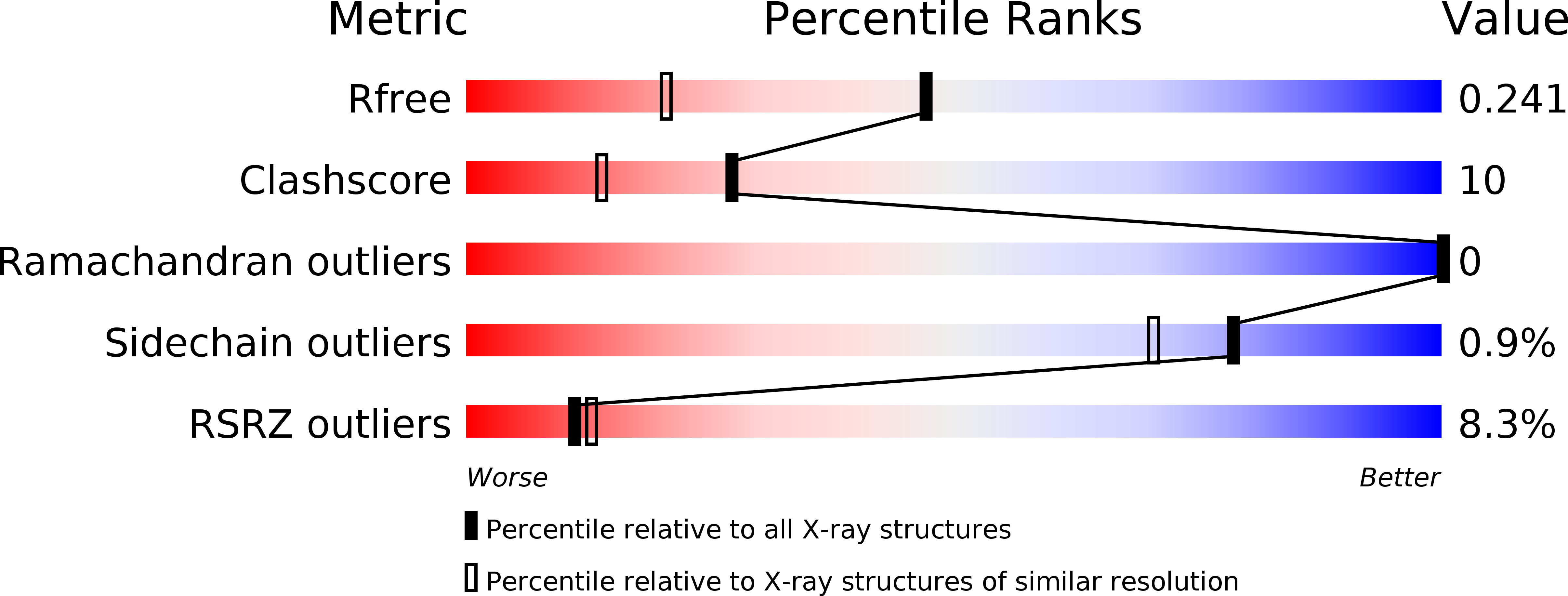
1ZPS image
Deposition Date 2005-05-17
Release Date 2005-08-30
Last Version Date 2024-02-14
Entry Detail
PDB ID:
1ZPS
Keywords:
Title:
Crystal structure of Methanobacterium thermoautotrophicum phosphoribosyl-AMP cyclohydrolase HisI
Biological Source:
Source Organism:
Host Organism:
Method Details:
Experimental Method:
Resolution:
1.70 Å
R-Value Free:
0.23
R-Value Work:
0.20
R-Value Observed:
0.20
Space Group:
P 21 21 21
Macromolecular Entities
Polymer Type:polypeptide(L)
Molecule:Phosphoribosyl-AMP cyclohydrolase
Gene (Uniprot):hisI
Chain IDs:A, B
Chain Length:138
Number of Molecules:2
Biological Source:Methanothermobacter thermautotrophicus
Primary Citation
Crystal Structure of Methanobacterium thermoautotrophicum Phosphoribosyl-AMP Cyclohydrolase HisI.
Biochemistry 44 10071 10080 (2005)
PMID: 16042384 DOI: 10.1021/bi050472w

Abstact

The metabolic pathway for histidine biosynthesis is interesting from an evolutionary perspective because of the diversity of gene organizations and protein structures involved. Hydrolysis of phosphoribosyl-AMP, the third step in the histidine biosynthetic pathway, is carried out by PR-AMP cyclohydrolase, the product of the hisI gene. The three-dimensional structure of PR-AMP cyclohydrolase from Methanobacterium thermoautotrophicum was solved and refined to 1.7 A resolution. The enzyme is a homodimer. The position of the Zn(2+)-binding site that is essential for catalysis was inferred from the positions of bound Cd(2+) ions, which were part of the crystallization medium. These metal binding sites include three cysteine ligands, two from one monomer and the third from the second monomer. The enzyme remains active when Cd(2+) is substituted for Zn(2+). The likely binding site for Mg(2+), also necessary for activity in a homologous cyclohydrolase, was also inferred from Cd(2+) positions and is comprised of aspartic acid side chains. The putative substrate-binding cleft is formed at the interface between the two monomers of the dimer. This fact, combined with the localization of the Zn(2+)-binding site, indicates that the enzyme is an obligate dimer.

Legend

Protein

Chemical

Disease

Primary Citation of related structures