1ZPD image
Deposition Date 1998-04-17
Release Date 1999-02-02
Last Version Date 2024-05-22
Entry Detail
PDB ID:
1ZPD
Title:
PYRUVATE DECARBOXYLASE FROM ZYMOMONAS MOBILIS
Biological Source:
Source Organism:
Host Organism:
Method Details:
Experimental Method:
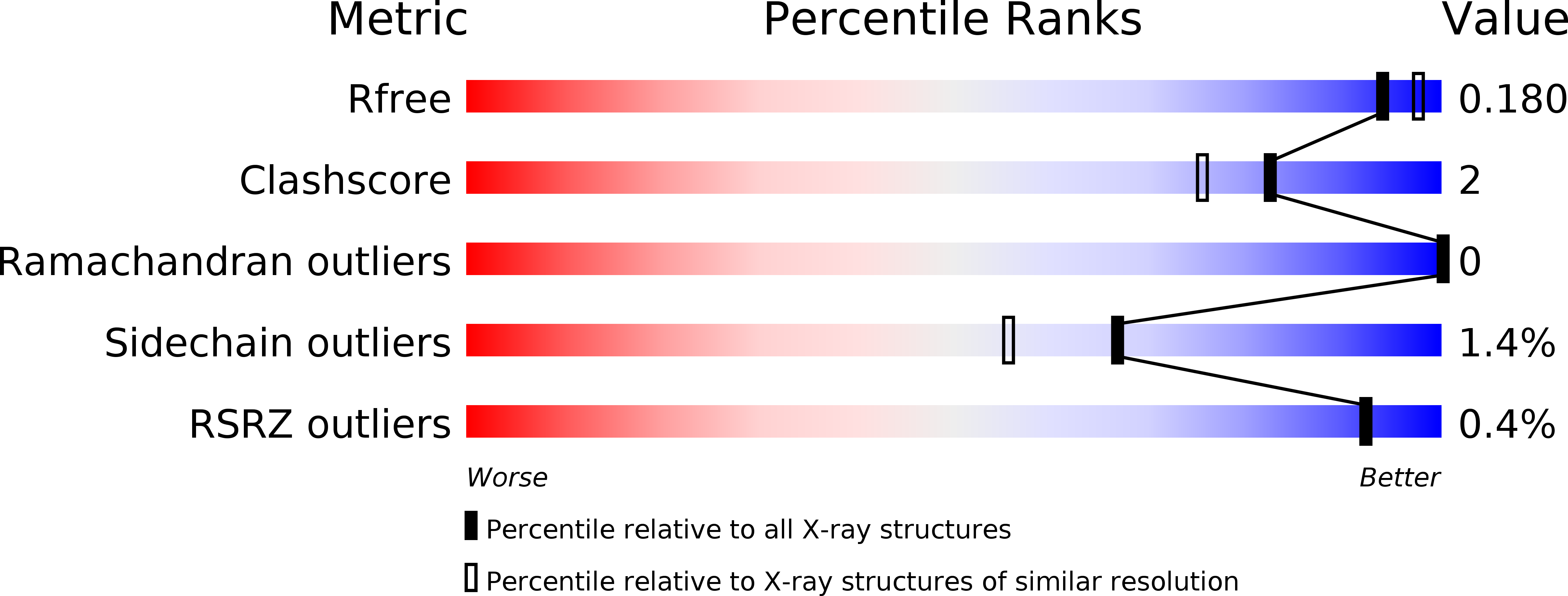
Resolution:
1.86 Å
R-Value Free:
0.19
R-Value Work:
0.16
Space Group:
P 1
Macromolecular Entities
Polymer Type:polypeptide(L)
Molecule:PYRUVATE DECARBOXYLASE
Gene (Uniprot):pdc
Chain IDs:A, B, C (auth: E), D (auth: F)
Chain Length:568
Number of Molecules:4
Biological Source:Zymomonas mobilis
Primary Citation
High resolution crystal structure of pyruvate decarboxylase from Zymomonas mobilis. Implications for substrate activation in pyruvate decarboxylases.
J.Biol.Chem. 273 20196 20204 (1998)
PMID: 9685367 DOI: 10.1074/jbc.273.32.20196

Abstact

The crystal structure of tetrameric pyruvate decarboxylase from Zymomonas mobilis has been determined at 1.9 A resolution and refined to a crystallographic R-factor of 16.2% and Rfree of 19.7%. The subunit consists of three domains, all of the alpha/beta type. Two of the subunits form a tight dimer with an extensive interface area. The thiamin diphosphate binding site is located at the subunit-subunit interface, and the cofactor, bound in the V conformation, interacts with residues from the N-terminal domain of one subunit and the C-terminal domain of the second subunit. The 2-fold symmetry generates the second thiamin diphosphate binding site in the dimer. Two of the dimers form a tightly packed tetramer with pseudo 222 symmetry. The interface area between the dimers is much larger in pyruvate decarboxylase from Z. mobilis than in the yeast enzyme, and structural differences in these parts result in a completely different packing of the subunits in the two enzymes. In contrast to other pyruvate decarboxylases, the enzyme from Z. mobilis is not subject to allosteric activation by the substrate. The tight packing of the dimers in the tetramer prevents large rearrangements in the quaternary structure as seen in the yeast enzyme and locks the enzyme in an activated conformation. The architecture of the cofactor binding site and the active site is similar in the two enzymes. However, the x-ray analysis reveals subtle but significant structural differences in the active site that might be responsible for variations in the biochemical properties in these enzymes.

Legend

Protein

Chemical

Disease

Primary Citation of related structures