1ZNN image
Deposition Date 2005-05-11
Release Date 2005-05-24
Last Version Date 2024-02-14
Entry Detail
PDB ID:
1ZNN
Title:
Structure of the synthase subunit of PLP synthase
Biological Source:
Source Organism:
Method Details:
Experimental Method:
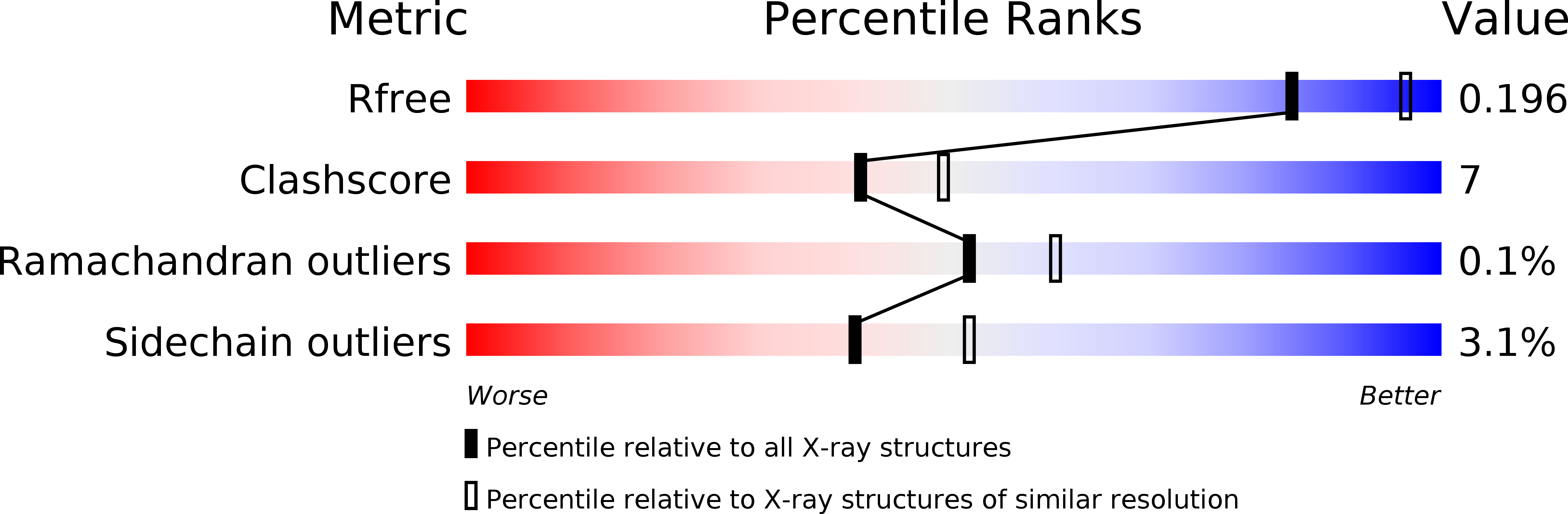
Resolution:
2.20 Å
R-Value Free:
0.19
R-Value Work:
0.15
R-Value Observed:
0.16
Space Group:
C 2 2 2
Macromolecular Entities
Polymer Type:polypeptide(L)
Molecule:PLP SYNTHASE
Chain IDs:A, B, C, D, E, F
Chain Length:325
Number of Molecules:6
Biological Source:Geobacillus stearothermophilus
Primary Citation
A New Arrangement of (beta/alpha)8 Barrels in the Synthase Subunit of PLP Synthase.
J.Biol.Chem. 280 27914 27923 (2005)
PMID: 15911615 DOI: 10.1074/jbc.M503642200

Abstact

Pyridoxal 5'-phosphate (PLP, vitamin B6), a cofactor in many enzymatic reactions, has two distinct biosynthetic routes, which do not coexist in any organism. Two proteins, known as PdxS and PdxT, together form a PLP synthase in plants, fungi, archaea, and some eubacteria. PLP synthase is a heteromeric glutamine amidotransferase in which PdxT produces ammonia from glutamine and PdxS combines ammonia with five- and three-carbon phosphosugars to form PLP. In the 2.2-A crystal structure, PdxS is a cylindrical dodecamer of subunits having the classic (beta/alpha)8 barrel fold. PdxS subunits form two hexameric rings with the active sites positioned on the inside. The hexamer and dodecamer forms coexist in solution. A novel phosphate-binding site is suggested by bound sulfate. The sulfate and another bound molecule, methyl pentanediol, were used to model the substrate ribulose 5-phosphate, and to propose catalytic roles for residues in the active site. The distribution of conserved surfaces in the PdxS dodecamer was used to predict a docking site for the glutaminase partner, PdxT.

Legend

Protein

Chemical

Disease

Primary Citation of related structures