Deposition Date
1989-09-25
Release Date
1991-07-15
Last Version Date
2024-11-20
Entry Detail
PDB ID:
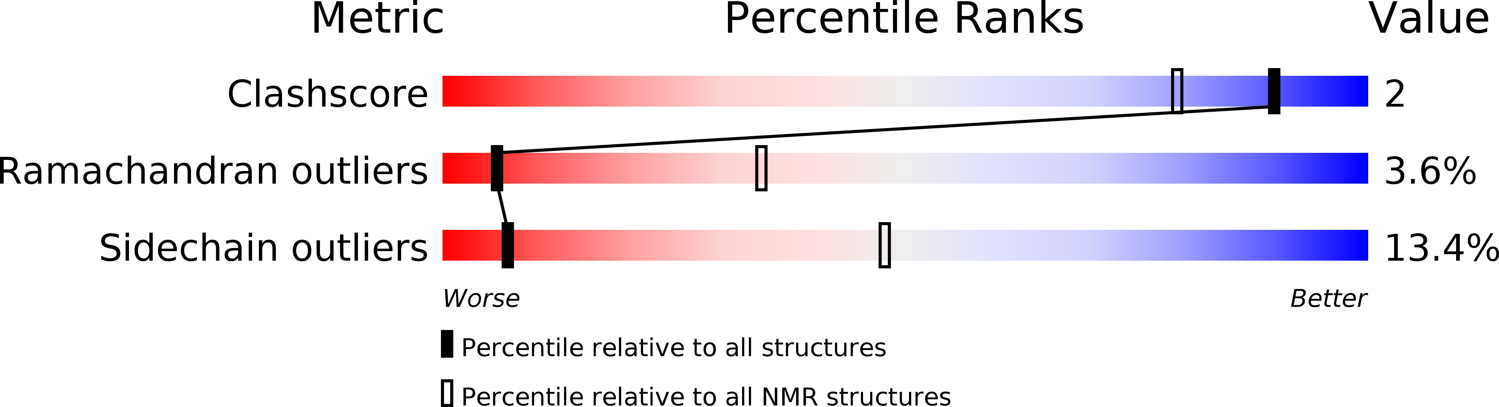
1ZNF
Keywords:
Title:
THREE-DIMENSIONAL SOLUTION STRUCTURE OF A SINGLE ZINC FINGER DNA-BINDING DOMAIN
Biological Source:
Source Organism(s):
Xenopus laevis (Taxon ID: 8355)
Method Details:
Experimental Method:
Conformers Submitted:
37