1ZKW image
Deposition Date 2005-05-04
Release Date 2005-06-28
Last Version Date 2023-08-23
Entry Detail
PDB ID:
1ZKW
Keywords:
Title:
Crystal structure of Arg347Ala mutant of botulinum neurotoxin E catalytic domain
Biological Source:
Source Organism:
Method Details:
Experimental Method:
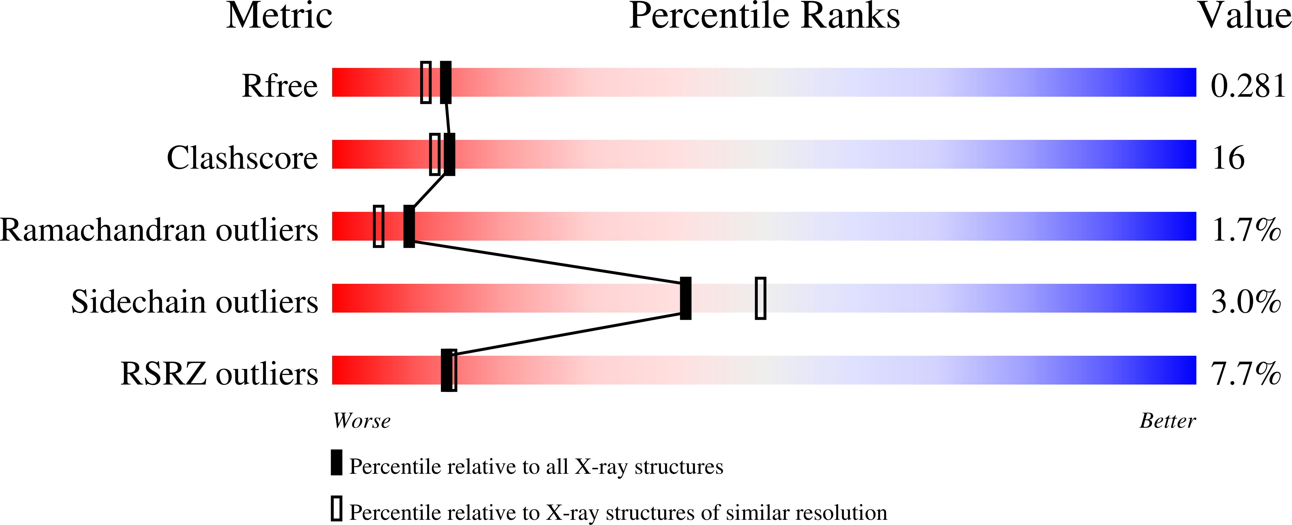
Resolution:
2.17 Å
R-Value Free:
0.28
R-Value Work:
0.23
Space Group:
P 21 21 2
Macromolecular Entities
Polymer Type:polypeptide(L)
Molecule:botulinum neurotoxin type E
Gene (Uniprot):botE
Mutations:R347A
Chain IDs:A, B
Chain Length:420
Number of Molecules:2
Biological Source:Clostridium botulinum
Primary Citation
Analysis of Active Site Residues of Botulinum Neurotoxin E by Mutational, Functional, and Structural Studies: Glu335Gln Is an Apoenzyme.
Biochemistry 44 8291 8302 (2005)
PMID: 15938619 DOI: 10.1021/bi050253a

Abstact

Clostridial neurotoxins comprising the seven serotypes of botulinum neurotoxins and tetanus neurotoxin are the most potent toxins known to humans. Their potency coupled with their specificity and selectivity underscores the importance in understanding their mechanism of action in order to develop a strategy for designing counter measures against them. To develop an effective vaccine against the toxin, it is imperative to achieve an inactive form of the protein which preserves the overall conformation and immunogenicity. Inactive mutants can be achieved either by targeting active site residues or by modifying the surface charges farther away from the active site. The latter affects the long-range forces such as electrostatic potentials in a subtle way without disturbing the structural integrity of the toxin causing some drastic changes in the activity/environment. Here we report structural and biochemical analysis on several mutations on Clostridium botulinum neurotoxin type E light chain with at least two producing dramatic effects: Glu335Gln causes the toxin to transform into a persistent apoenzyme devoid of zinc, and Tyr350Ala has no hydrolytic activity. The structural analysis of several mutants has led to a better understanding of the catalytic mechanism of this family of proteins. The residues forming the S1' subsite have been identified by comparing this structure with a thermolysin-inhibitor complex structure.

Legend

Protein

Chemical

Disease

Primary Citation of related structures