Deposition Date
2005-04-27
Release Date
2005-12-13
Last Version Date
2023-08-23
Entry Detail
PDB ID:
1ZJ2
Keywords:
Title:
Crystal Structure of Human Galactosyltransferase (GTB) Complexed with H type I Trisaccharide
Biological Source:
Source Organism(s):
Homo sapiens (Taxon ID: 9606)
Expression System(s):
Method Details:
Experimental Method:
Resolution:
1.69 Å
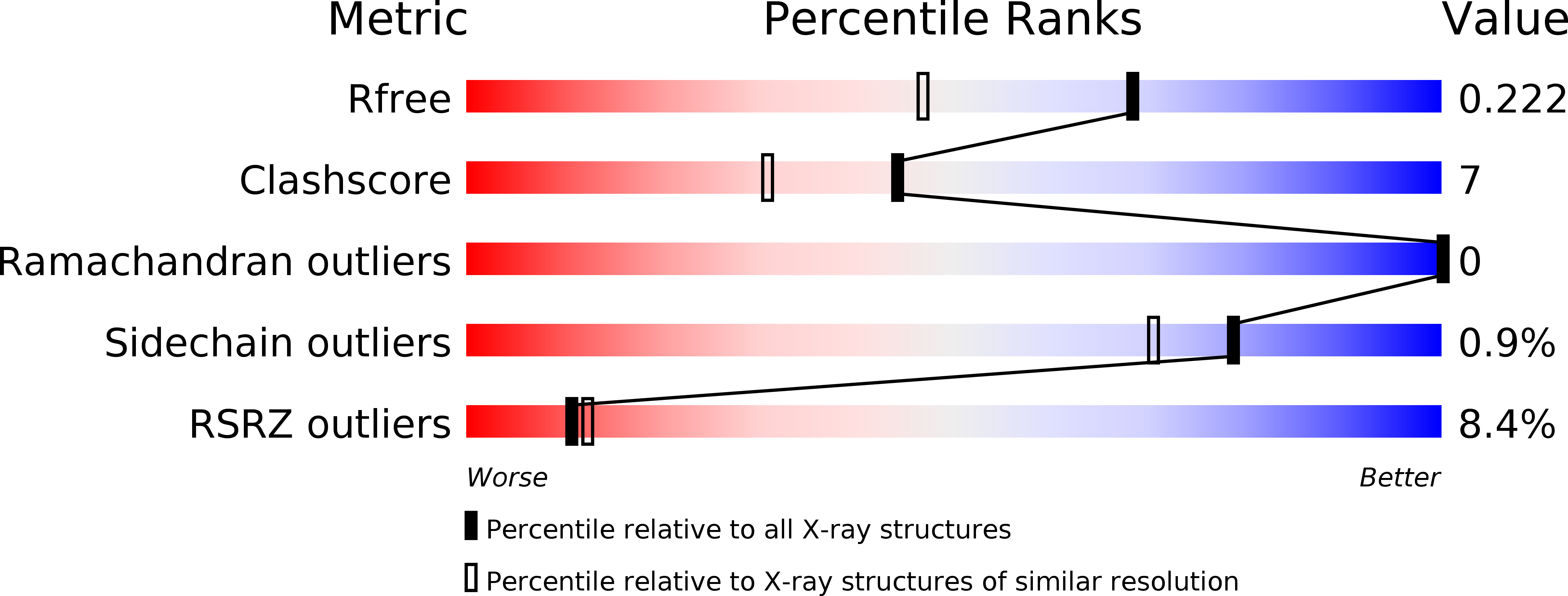
R-Value Free:
0.23
R-Value Work:
0.20
Space Group:
C 2 2 21