
Deposition Date
2005-04-27
Release Date
2005-07-05
Last Version Date
2023-10-25
Entry Detail
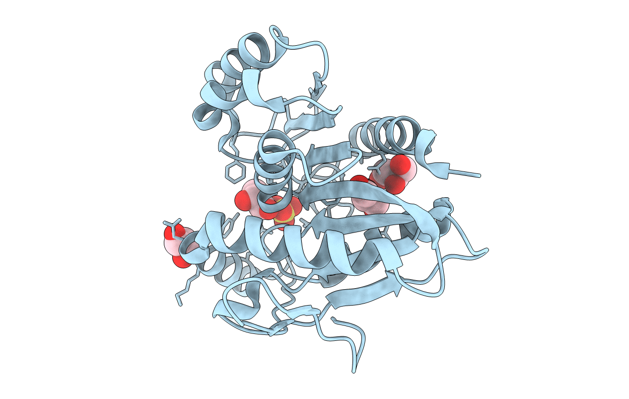
PDB ID:
1ZI9
Keywords:
Title:
Crystal Structure Analysis of the dienelactone hydrolase (E36D, C123S) mutant- 1.5 A
Biological Source:
Source Organism(s):
Pseudomonas putida (Taxon ID: 303)
Expression System(s):
Method Details:
Experimental Method:
Resolution:
1.50 Å
R-Value Free:
0.19
R-Value Work:
0.17
R-Value Observed:
0.17
Space Group:
P 21 21 21
