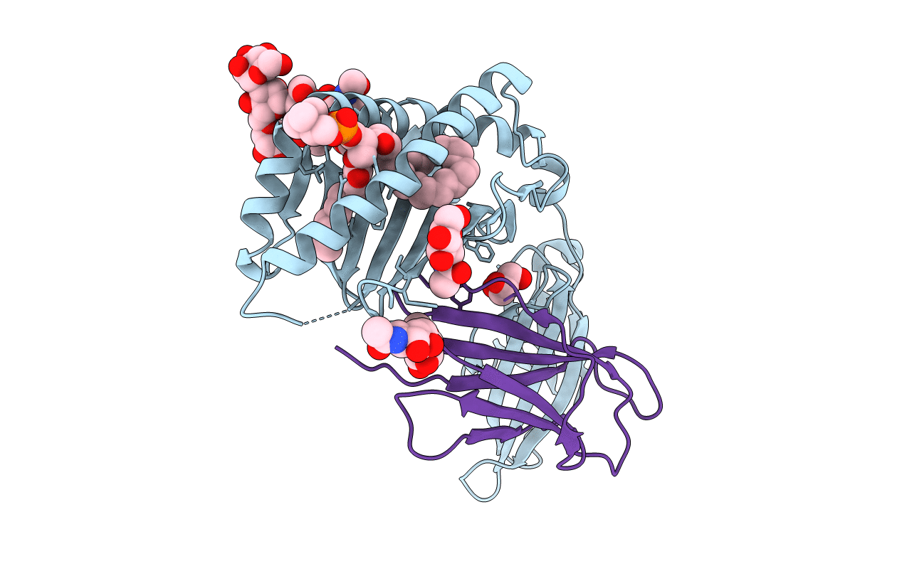
1ZHN image
Deposition Date 2005-04-26
Release Date 2005-07-19
Last Version Date 2024-10-30
Entry Detail
PDB ID:
1ZHN
Title:
Crystal Structure of mouse CD1d bound to the self ligand phosphatidylcholine
Biological Source:
Source Organism:
Mus musculus (Taxon ID: 10090)
Method Details:
Experimental Method:
Resolution:
2.80 Å
R-Value Free:
0.28
R-Value Work:
0.21
R-Value Observed:
0.22
Space Group:
P 21 21 21
Macromolecular Entities
Polymer Type:polypeptide(L)
Molecule:CD1d1 antigen
Chain IDs:A
Chain Length:273
Number of Molecules:1
Biological Source:Mus musculus
Polymer Type:polypeptide(L)
Molecule:beta-2-microglobulin
Gene (Uniprot):B2m
Chain IDs:B
Chain Length:99
Number of Molecules:1
Biological Source:Mus musculus
Modified Residue
Compound ID Chain ID Parent Comp ID Details 2D Image
ASN A ASN GLYCOSYLATION SITE
Primary Citation
Crystal structure of mouse CD1d bound to the self ligand phosphatidylcholine: a molecular basis for NKT cell activation
J.Immunol. 175 977 984 (2005)
PMID: 16002697

Abstact

NKT cells are immunoregulatory lymphocytes whose activation is triggered by the recognition of lipid Ags in the context of the CD1d molecules by the TCR. In this study we present the crystal structure to 2.8 A of mouse CD1d bound to phosphatidylcholine. The interactions between the ligand acyl chains and the CD1d molecule define the structural and chemical requirements for the binding of lipid Ags to CD1d. The orientation of the polar headgroup toward the C terminus of the alpha1 helix provides a rationale for the structural basis for the observed Valpha chain bias in invariant NKT cells. The contribution of the ligand to the protein surface suggests a likely mode of recognition of lipid Ags by the NKT cell TCR.

Legend

Protein

Chemical

Disease

Primary Citation of related structures