1ZH1 image
Deposition Date 2005-04-22
Release Date 2005-05-24
Last Version Date 2024-10-16
Entry Detail
PDB ID:
1ZH1
Title:
Structure of the zinc-binding domain of HCV NS5A
Biological Source:
Source Organism:
Method Details:
Experimental Method:
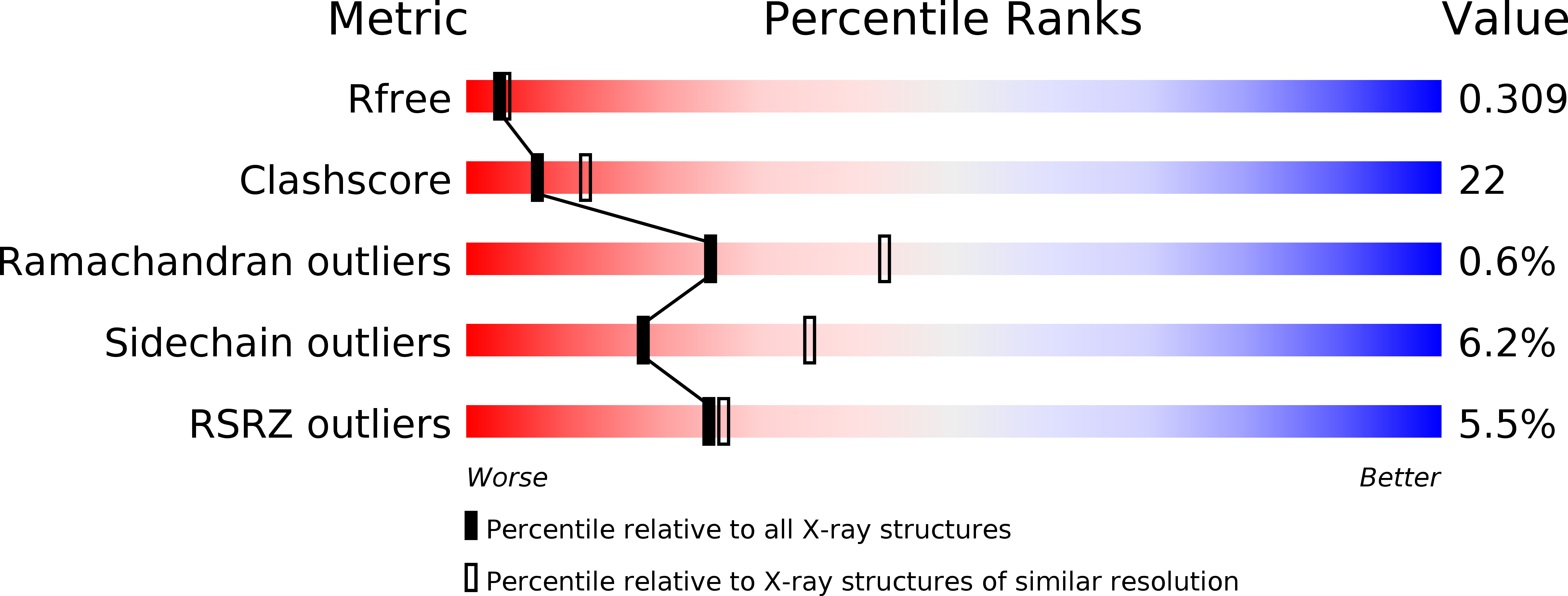
Resolution:
2.50 Å
R-Value Free:
0.28
R-Value Work:
0.22
Space Group:
P 41 2 2
Macromolecular Entities
Polymer Type:polypeptide(L)
Molecule:non-structural polyprotein
Chain IDs:A, B
Chain Length:163
Number of Molecules:2
Biological Source:Hepatitis C virus
Ligand Molecules
Primary Citation
Structure of the zinc-binding domain of an essential component of the hepatitis C virus replicase.
Nature 435 374 379 (2005)
PMID: 15902263 DOI: 10.1038/nature03580

Abstact

Hepatitis C virus (HCV) is a human pathogen affecting nearly 3% of the world's population. Chronic infections can lead to cirrhosis and liver cancer. The RNA replication machine of HCV is a multi-subunit membrane-associated complex. The non-structural protein NS5A is an active component of HCV replicase, as well as a pivotal regulator of replication and a modulator of cellular processes ranging from innate immunity to dysregulated cell growth. NS5A is a large phosphoprotein (56-58 kDa) with an amphipathic alpha-helix at its amino terminus that promotes membrane association. After this helix region, NS5A is organized into three domains. The N-terminal domain (domain I) coordinates a single zinc atom per protein molecule. Mutations disrupting either the membrane anchor or zinc binding of NS5A are lethal for RNA replication. However, probing the role of NS5A in replication has been hampered by a lack of structural information about this multifunctional protein. Here we report the structure of NS5A domain I at 2.5-A resolution, which contains a novel fold, a new zinc-coordination motif and a disulphide bond. We use molecular surface analysis to suggest the location of protein-, RNA- and membrane-interaction sites.

Legend

Protein

Chemical

Disease

Primary Citation of related structures