1ZGR image
Deposition Date 2005-04-22
Release Date 2005-10-11
Last Version Date 2024-03-13
Entry Detail
PDB ID:
1ZGR
Title:
Crystal structure of the Parkia platycephala seed lectin
Biological Source:
Source Organism:
Method Details:
Experimental Method:
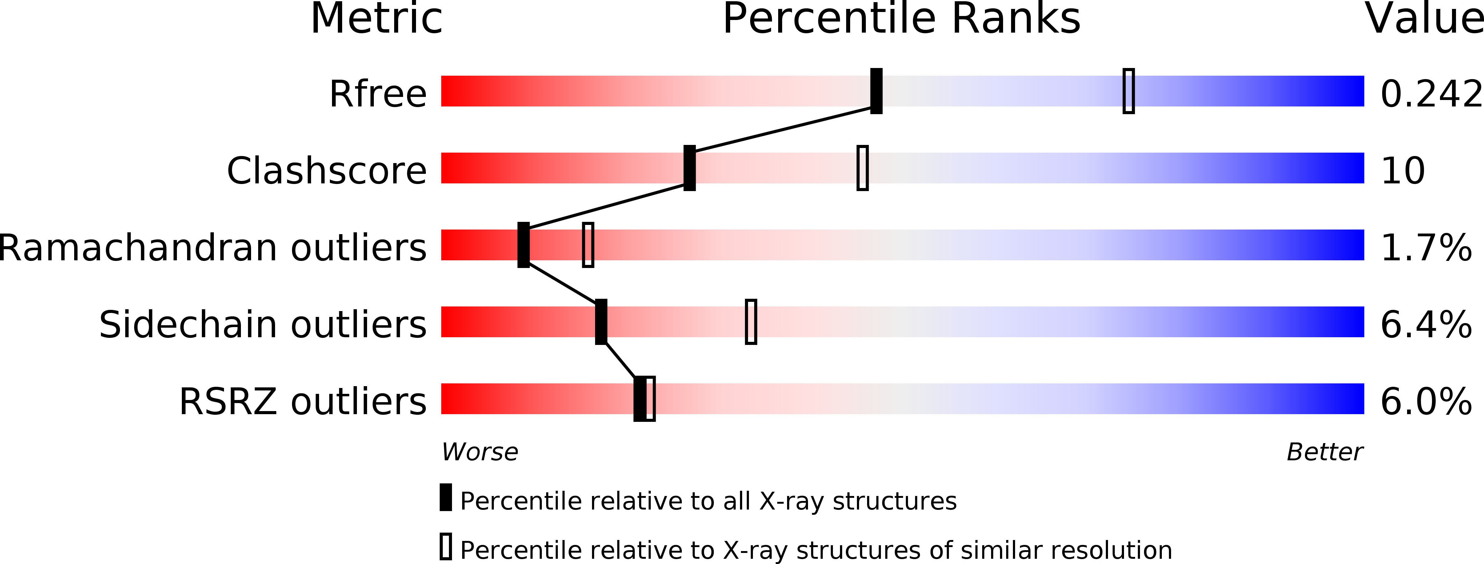
Resolution:
2.50 Å
R-Value Free:
0.24
R-Value Work:
0.21
R-Value Observed:
0.21
Space Group:
P 21 21 21
Macromolecular Entities
Polymer Type:polypeptide(L)
Molecule:Mannose/glucose-specific lectin
Chain IDs:A, B
Chain Length:447
Number of Molecules:2
Biological Source:Parkia platycephala
Primary Citation
The first crystal structure of a mimosoideae lectin reveals a novel quaternary arrangement of a widespread domain.
J.Mol.Biol. 353 574 583 (2005)
PMID: 16185708 DOI: 10.1016/j.jmb.2005.08.055

Abstact

The crystal structures of the apo and mannose-bound Parkia platycephala seed lectin represent the first structure of a Mimosoideae lectin and a novel circular arrangement of beta-prism domains, and highlight the adaptability of the beta-prism fold as a building block in the evolution of plant lectins. The P.platycephala lectin is a dimer both in solution and in the crystals. Mannose binding to each of the three homologous carbohydrate-recognition domains of the lectin occurs through different modes, and restrains the flexibility of surface-exposed loops and residues involved in carbohydrate recognition. The planar array of carbohydrate-binding sites on the rim of the toroid-shaped structure of the P.platycephala lectin dimer immediately suggests a mechanism to promote multivalent interactions leading to cross-linking of carbohydrate ligands as part of the host strategy against phytopredators and pathogens. The cyclic structure of the P.platycephala lectin points to the convergent evolution of a structural principle for the construction of lectins involved in host defense or in attacking other organisms.

Legend

Protein

Chemical

Disease

Primary Citation of related structures