1ZES image
Deposition Date 2005-04-19
Release Date 2005-09-20
Last Version Date 2024-02-14
Entry Detail
PDB ID:
1ZES
Title:
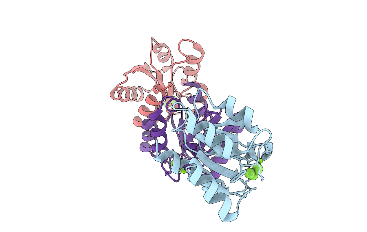
BeF3- activated PhoB receiver domain
Biological Source:
Source Organism:
Escherichia coli (Taxon ID: 562)
Host Organism:
Method Details:
Experimental Method:
Resolution:
1.90 Å
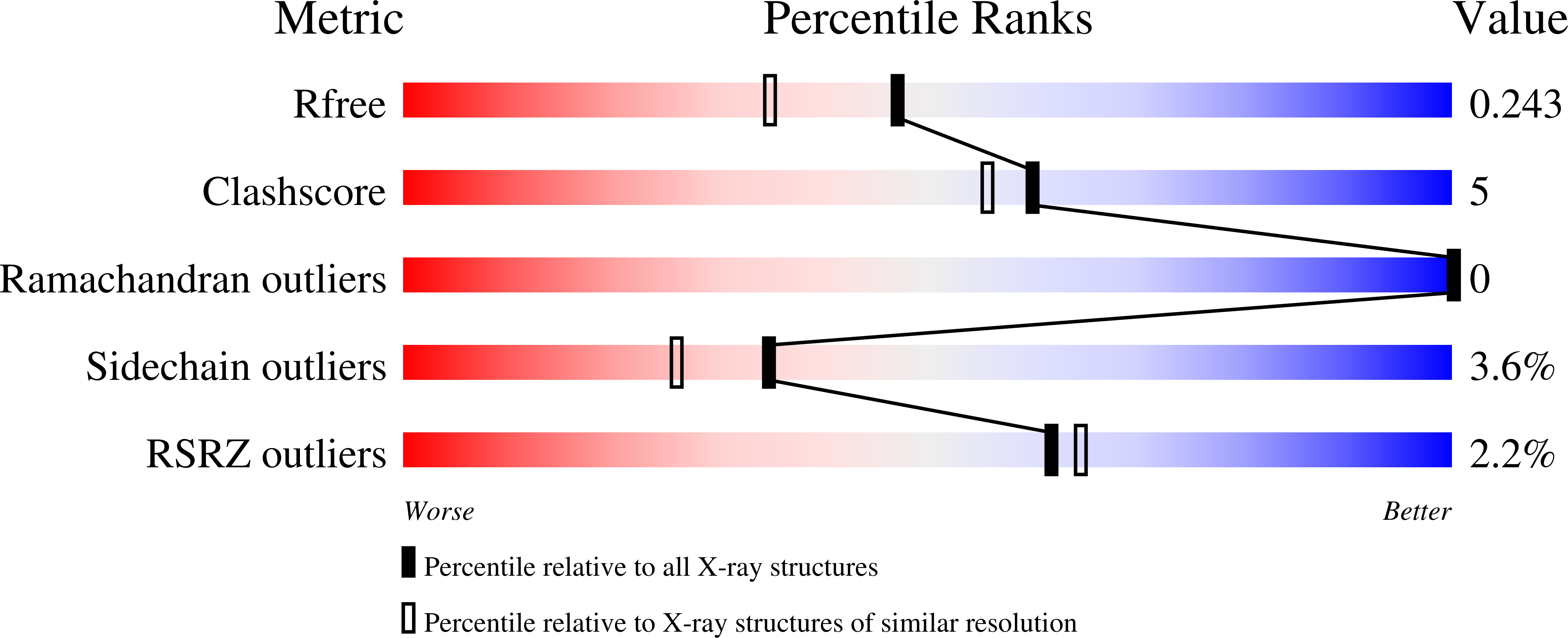
R-Value Free:
0.24
R-Value Work:
0.18
R-Value Observed:
0.19
Space Group:
P 43 21 2
Macromolecular Entities
Polymer Type:polypeptide(L)
Molecule:Phosphate regulon transcriptional regulatory protein phoB
Mutations:P125Q
Chain IDs:A, B, C
Chain Length:125
Number of Molecules:3
Biological Source:Escherichia coli
Primary Citation
Mechanism of Activation for Transcription Factor PhoB Suggested by Different Modes of Dimerization in the Inactive and Active States.
Structure 13 1353 1363 (2005)
PMID: 16154092 DOI: 10.1016/j.str.2005.06.006

Abstact

Response regulators (RRs), which undergo phosphorylation/dephosphorylation at aspartate residues, are highly prevalent in bacterial signal transduction. RRs typically contain an N-terminal receiver domain that regulates the activities of a C-terminal DNA binding domain in a phosphorylation-dependent manner. We present crystallography and solution NMR data for the receiver domain of Escherichia coli PhoB which show distinct 2-fold symmetric dimers in the inactive and active states. These structures, together with the previously determined structure of the C-terminal domain of PhoB bound to DNA, define the conformation of the active transcription factor and provide a model for the mechanism of activation in the OmpR/PhoB subfamily, the largest group of RRs. In the active state, the receiver domains dimerize with 2-fold rotational symmetry using their alpha4-beta5-alpha5 faces, while the effector domains bind to DNA direct repeats with tandem symmetry, implying a loss of intramolecular interactions.

Legend

Protein

Chemical

Disease

Primary Citation of related structures