Deposition Date
2005-04-19
Release Date
2005-08-02
Last Version Date
2024-02-14
Entry Detail
PDB ID:
1ZEQ
Keywords:
Title:
1.5 A Structure of apo-CusF residues 6-88 from Escherichia coli
Biological Source:
Source Organism(s):
Escherichia coli (Taxon ID: 562)
Expression System(s):
Method Details:
Experimental Method:
Resolution:
1.50 Å
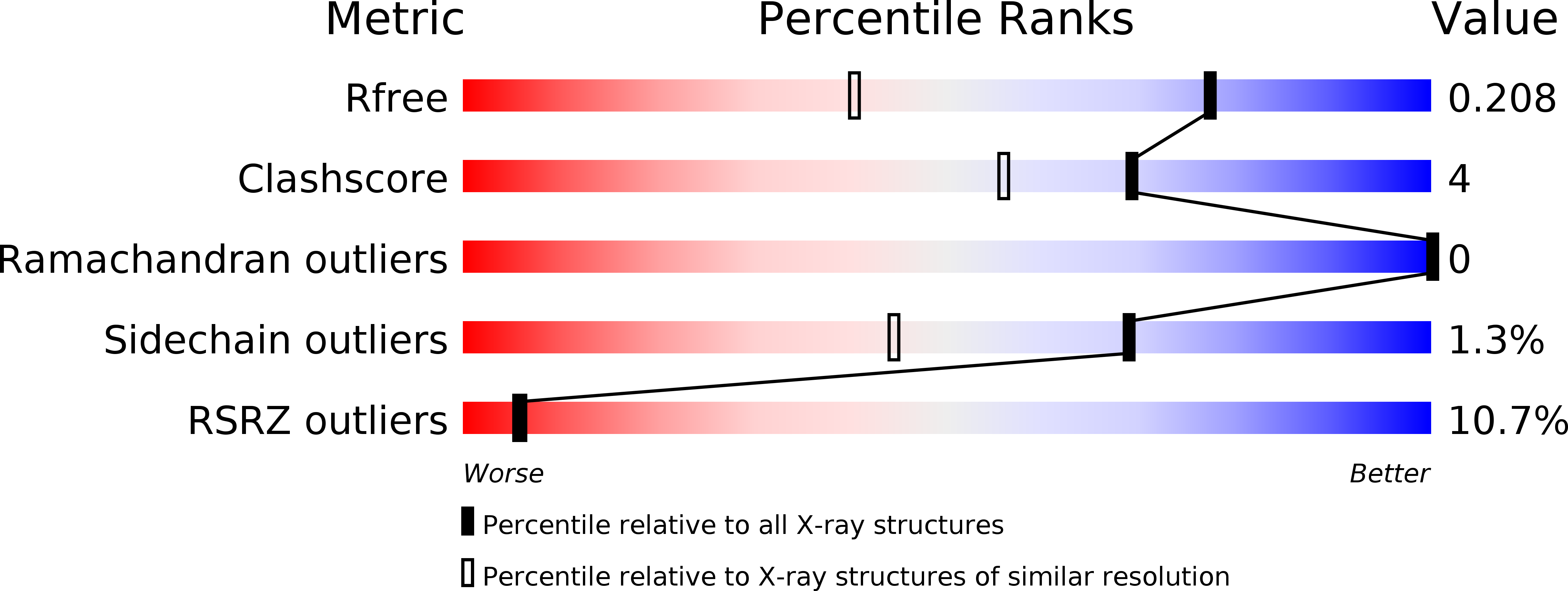
R-Value Free:
0.20
R-Value Work:
0.18
R-Value Observed:
0.18
Space Group:
P 21 21 21