1ZDU image
Deposition Date 2005-04-14
Release Date 2005-05-24
Last Version Date 2024-02-14
Entry Detail
PDB ID:
1ZDU
Keywords:
Title:
The Crystal Structure of Human Liver Receptor Homologue-1
Biological Source:
Source Organism:
Homo sapiens (Taxon ID: 9606)
(Taxon ID: )
Host Organism:
Method Details:
Experimental Method:
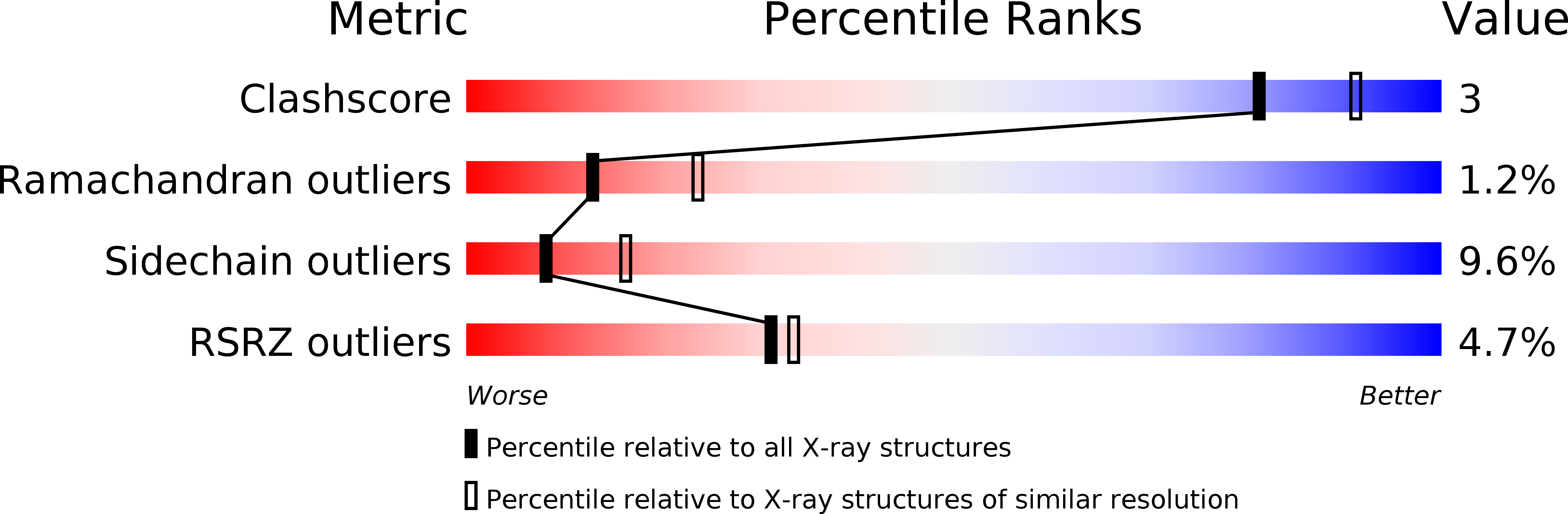
Resolution:
2.50 Å
R-Value Free:
0.28
R-Value Work:
0.23
R-Value Observed:
0.24
Space Group:
P 21 21 21
Macromolecular Entities
Polymer Type:polypeptide(L)
Molecule:Orphan nuclear receptor NR5A2
Gene (Uniprot):NR5A2
Chain IDs:A
Chain Length:245
Number of Molecules:1
Biological Source:Homo sapiens
Polymer Type:polypeptide(L)
Molecule:Nuclear receptor coactivator 2
Gene (Uniprot):NCOA2
Chain IDs:B (auth: P), C (auth: Q)
Chain Length:11
Number of Molecules:2
Biological Source:
Primary Citation
The crystal structures of human steroidogenic factor-1 and liver receptor homologue-1
Proc.Natl.Acad.Sci.USA 102 7505 7510 (2005)
PMID: 15897460 DOI: 10.1073/pnas.0409482102

Abstact

Steroidogenic factor-1 (SF-1) and liver receptor homologue-1 (LRH-1) belong to the fushi tarazu factor 1 subfamily of nuclear receptors. SF-1 is an essential factor for sex determination during development and regulates adrenal and gonadal steroidogenesis in the adult, whereas LRH-1 is a critical factor for development of endodermal tissues and regulates cholesterol and bile acid homeostasis. Regulatory ligands are unknown for SF-1 and LRH-1. A reported mouse LRH-1 structure revealed an empty pocket in a region commonly occupied by ligands in the structures of other nuclear receptors, and pocket-filling mutations did not alter the constitutive activity observed. Here we report the crystal structures of the putative ligand-binding domains of human SF-1 at 2.1-A resolution and human LRH-1 at 2.5-A resolution. Both structures bind a coactivator-derived peptide at the canonical activation-function surface, thus adopting the transcriptionally activating conformation. In human LRH-1, coactivator peptide binding also occurs to a second site. We discovered in both structures a phospholipid molecule bound in a pocket of the putative ligand-binding domain. MS analysis of the protein samples used for crystallization indicated that the two proteins associate with a range of phospholipids. Mutations of the pocket-lining residues reduced the transcriptional activities of SF-1 and LRH-1 in mammalian cell transfection assays without affecting their expression levels. These results suggest that human SF-1 and LRH-1 may be ligand-binding receptors, although it remains to be seen if phospholipids or possibly other molecules regulate SF-1 or LRH-1 under physiological conditions.

Legend

Protein

Chemical

Disease

Primary Citation of related structures