Deposition Date
2005-04-09
Release Date
2005-12-06
Last Version Date
2024-05-22
Entry Detail
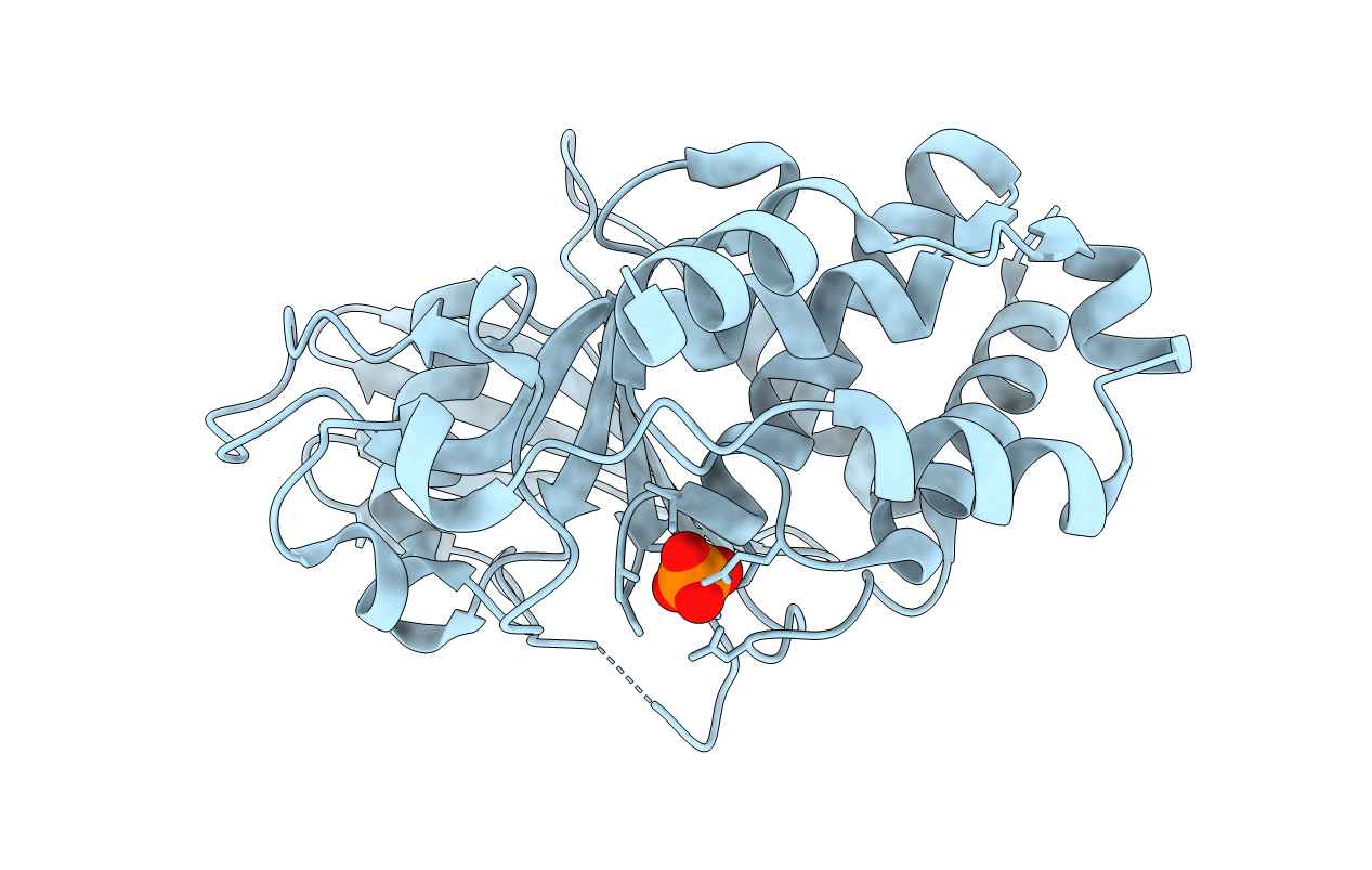
PDB ID:
1ZC0
Keywords:
Title:
Crystal structure of human hematopoietic tyrosine phosphatase (HePTP) catalytic domain
Biological Source:
Source Organism(s):
Homo sapiens (Taxon ID: 9606)
Expression System(s):
Method Details:
Experimental Method:
Resolution:
1.85 Å
R-Value Free:
0.18
R-Value Work:
0.16
R-Value Observed:
0.16
Space Group:
P 61