1ZB7 image
Deposition Date 2005-04-07
Release Date 2005-07-05
Last Version Date 2024-10-30
Entry Detail
PDB ID:
1ZB7
Keywords:
Title:
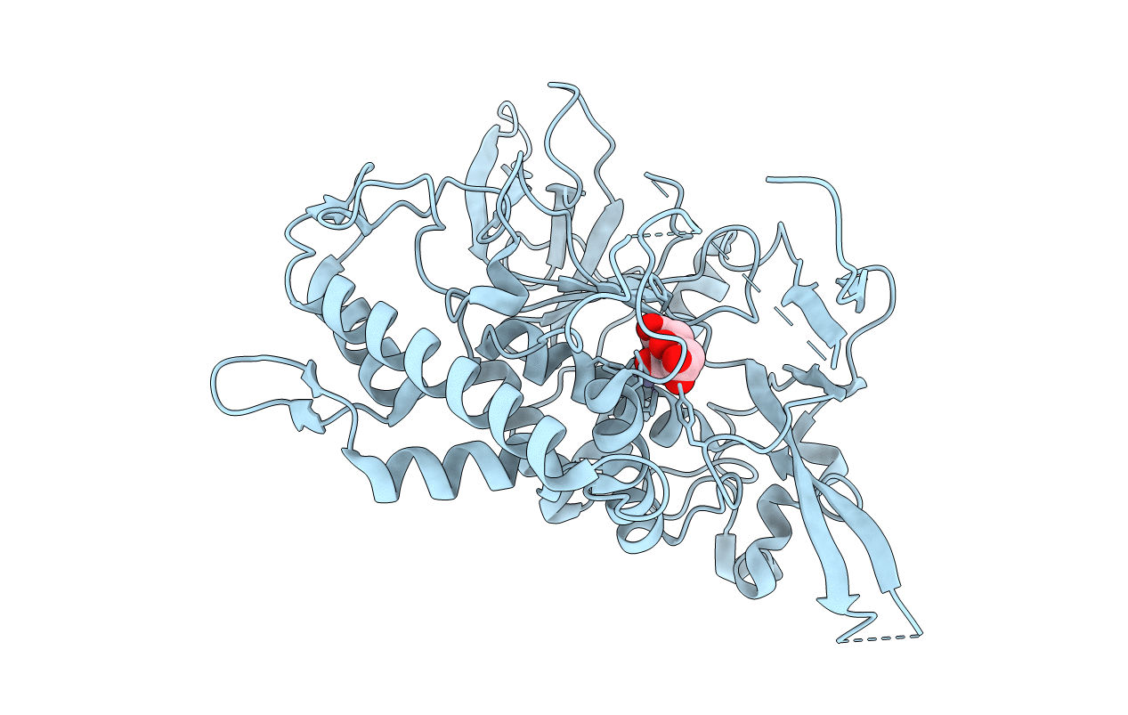
Crystal Structure of Botulinum Neurotoxin Type G Light Chain
Biological Source:
Source Organism:
Host Organism:
Method Details:
Experimental Method:
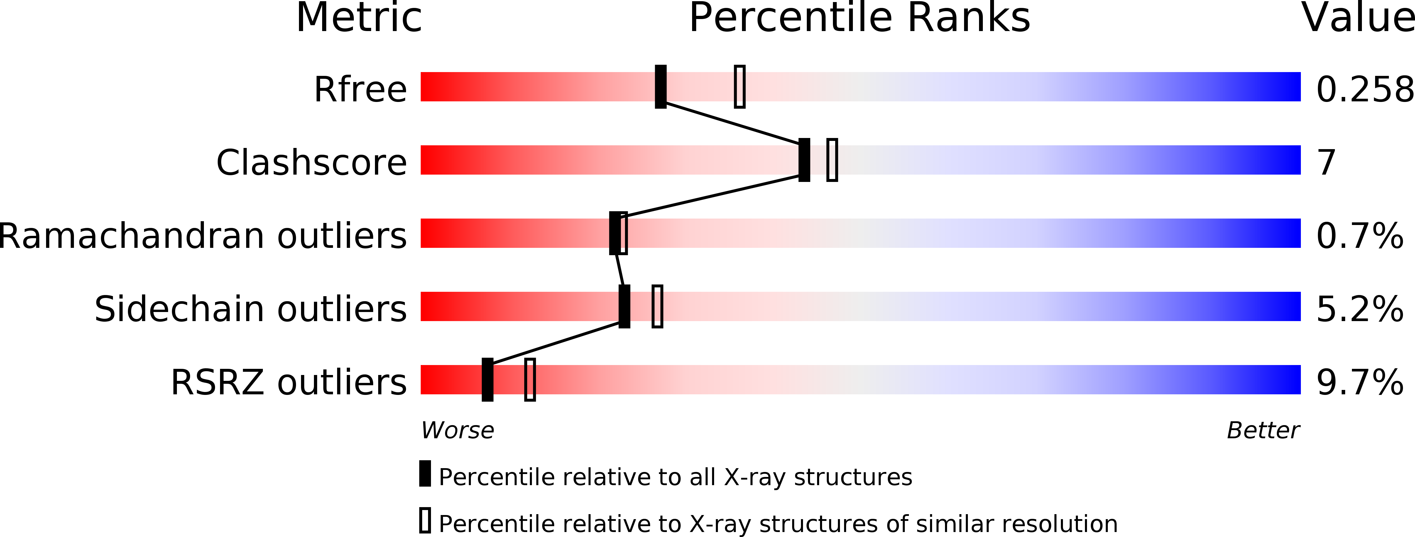
Resolution:
2.35 Å
R-Value Free:
0.22
R-Value Work:
0.17
R-Value Observed:
0.17
Space Group:
P 62 2 2
Macromolecular Entities
Structures with similar UniProt ID
Protein Blast
Polymer Type:polypeptide(L)
Molecule:neurotoxin
Gene (Uniprot):botG
Chain IDs:A
Chain Length:455
Number of Molecules:1
Biological Source:Clostridium botulinum
Primary Citation
Crystal structure of botulinum neurotoxin type g light chain: serotype divergence in substrate recognition
Biochemistry 44 9574 9580 (2005)
PMID: 16008342 DOI: 10.1021/bi0505924

Abstact

The seven serotypes (A-G) of botulinum neurotoxins (BoNTs) block neurotransmitter release through their specific proteolysis of one of the three proteins of the soluble N-ethylmaleimide-sensitive-factor attachment protein receptor (SNARE) complex. BoNTs have stringent substrate specificities that are unique for metalloprotease in that they require exceptionally long substrates (1). To understand the molecular reasons for the unique specificities of the BoNTs, we determined the crystal structure of the catalytic light chain (LC) of Clostridium botulinum neurotoxin type G (BoNT/G-LC) at 2.35 A resolution. The structure of BoNT/G-LC reveals a C-terminal beta-sheet that is critical for LC oligomerization and is unlike that seen in the other LC structures. Its structural comparison with thermolysin and the available pool of LC structures reveals important serotype differences that are likely to be involved in substrate recognition of the P1' residue. In addition, structural and sequence analyses have identified a potential exosite of BoNT/G-LC that recognizes a SNARE recognition motif of VAMP.

Legend

Protein

Chemical

Disease

Primary Citation of related structures