Deposition Date
2005-04-05
Release Date
2005-05-10
Last Version Date
2024-11-20
Entry Detail
PDB ID:
1ZA6
Keywords:
Title:
The structure of an antitumor CH2-domain-deleted humanized antibody
Biological Source:
Source Organism:
Homo sapiens (Taxon ID: 9606)
Host Organism:
Method Details:
Experimental Method:
Resolution:
2.80 Å
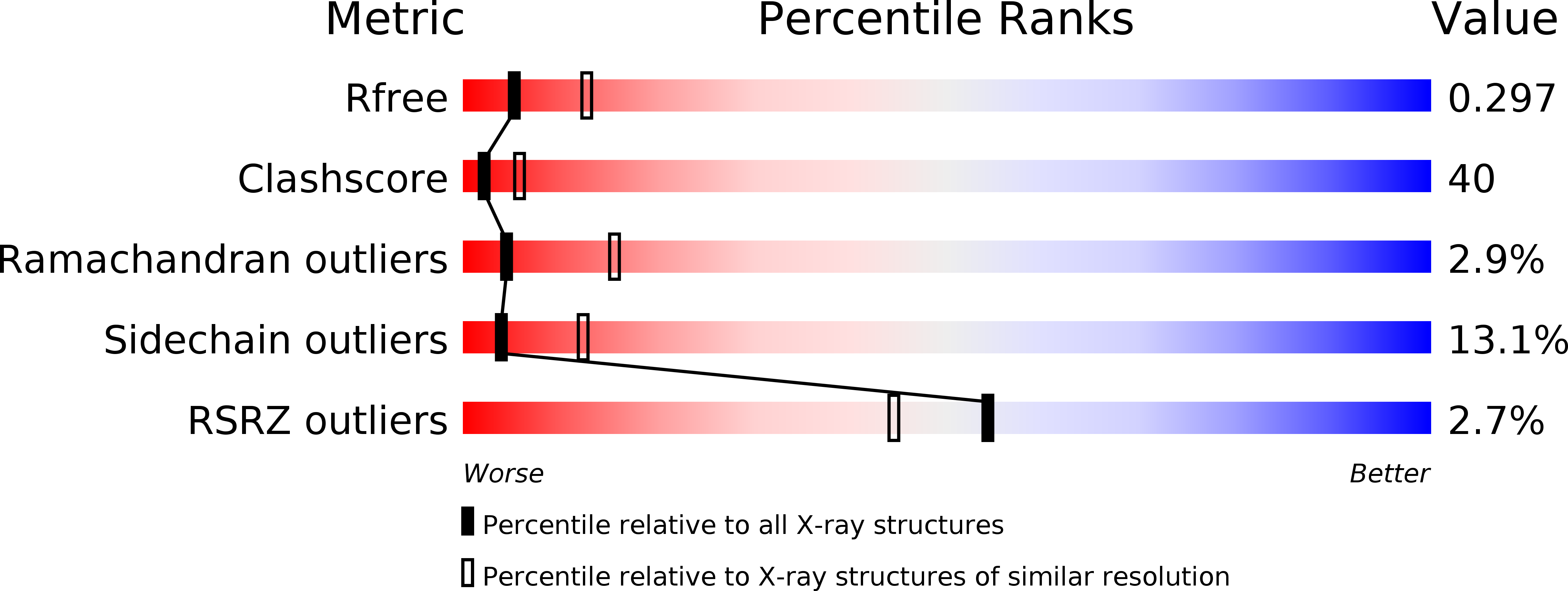
R-Value Free:
0.29
R-Value Work:
0.24
R-Value Observed:
0.24
Space Group:
P 21 21 2