Deposition Date
2005-04-03
Release Date
2005-07-19
Last Version Date
2024-11-20
Entry Detail
PDB ID:
1Z9L
Keywords:
Title:
1.7 Angstrom Crystal Structure of the Rat VAP-A MSP Homology Domain
Biological Source:
Source Organism(s):
Rattus norvegicus (Taxon ID: 10116)
Expression System(s):
Method Details:
Experimental Method:
Resolution:
1.70 Å
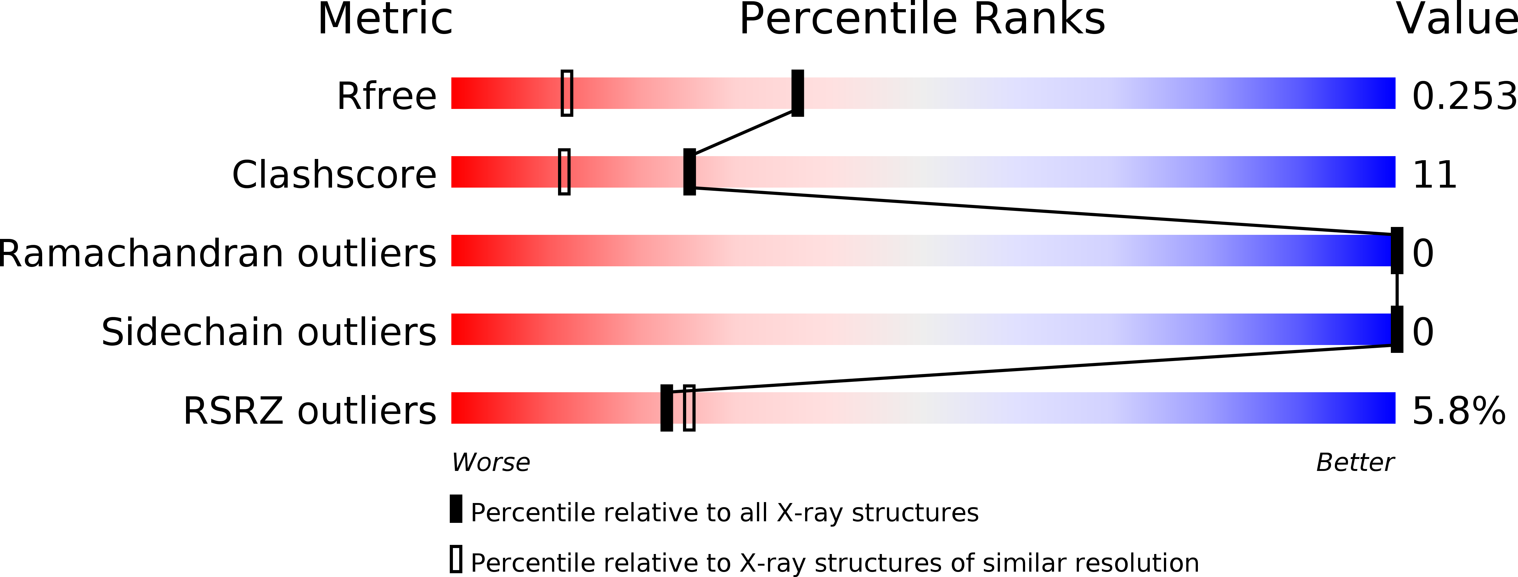
R-Value Free:
0.25
R-Value Work:
0.22
R-Value Observed:
0.22
Space Group:
P 42 21 2