1Z9K image
Deposition Date 2005-04-02
Release Date 2005-06-07
Last Version Date 2023-08-23
Entry Detail
PDB ID:
1Z9K
Keywords:
Title:
Photosynthetic Reaction Center from Rhodobacter sphaeroides
Biological Source:
Source Organism:
Method Details:
Experimental Method:
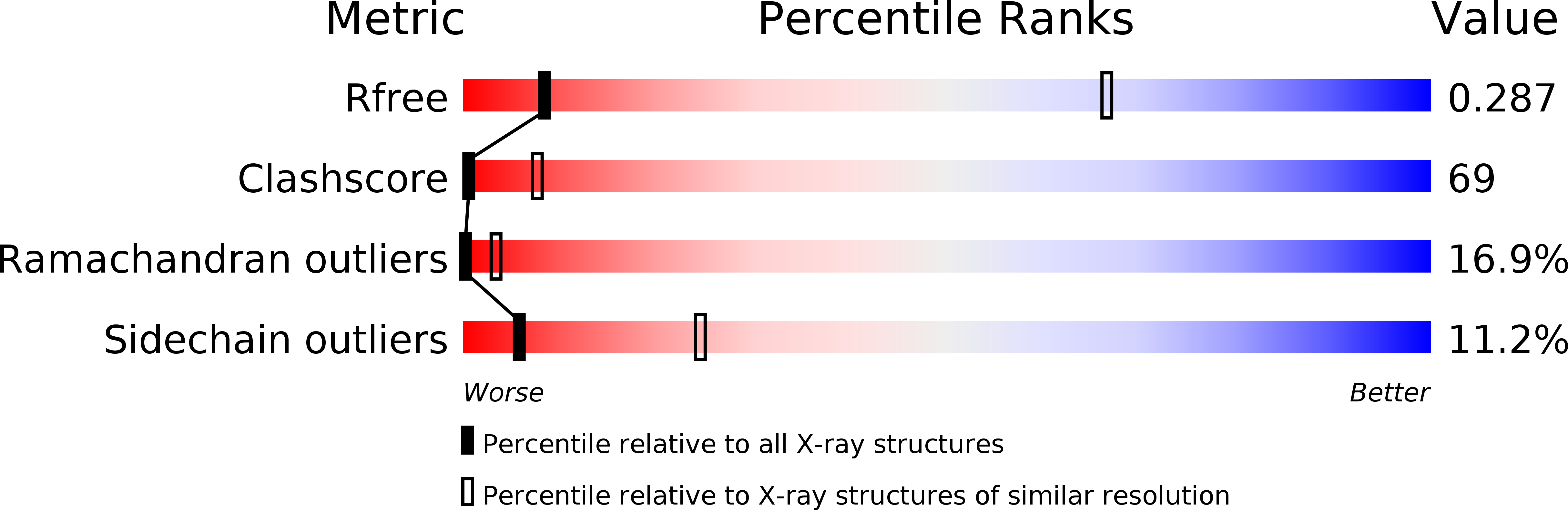
Resolution:
4.60 Å
R-Value Free:
0.33
R-Value Work:
0.33
R-Value Observed:
0.34
Space Group:
P 42 2 2
Macromolecular Entities
Polymer Type:polypeptide(L)
Molecule:Reaction center protein L chain
Chain IDs:A
Chain Length:281
Number of Molecules:1
Biological Source:Rhodobacter sphaeroides
Polymer Type:polypeptide(L)
Molecule:Reaction center protein M chain
Chain IDs:B
Chain Length:307
Number of Molecules:1
Biological Source:Rhodobacter sphaeroides
Polymer Type:polypeptide(L)
Molecule:Reaction center protein H chain
Chain IDs:C
Chain Length:260
Number of Molecules:1
Biological Source:Rhodobacter sphaeroides
Primary Citation
Design of a Redox-Linked Active Metal Site: Manganese Bound to Bacterial Reaction Centers at a Site Resembling That of Photosystem II
Biochemistry 44 7389 7394 (2005)
PMID: 15895982 DOI: 10.1021/bi050377n

Abstact

Metals bound to proteins perform a number of crucial biological reactions, including the oxidation of water by a manganese cluster in photosystem II. Although evolutionarily related to photosystem II, bacterial reaction centers lack both a strong oxidant and a manganese cluster for mediating the multielectron and proton transfer needed for water oxidation. In this study, carboxylate residues were introduced by mutagenesis into highly oxidizing reaction centers at a site homologous to the manganese-binding site of photosystem II. In the presence of manganese, light-minus-dark difference optical spectra of reaction centers from the mutants showed a lack of the oxidized bacteriochlorophyll dimer, while the reduced primary quinone was still present, demonstrating that manganese was serving as a secondary electron donor. On the basis of these steady-state optical measurements, the mutant with the highest-affinity site had a dissociation constant of approximately 1 microM. For the highest-affinity mutant, a first-order rate with a lifetime of 12 ms was observed for the reduction of the oxidized bacteriochlorophyll dimer by the bound manganese upon exposure to light. The dependence of the amplitude of this component on manganese concentration yielded a dissociation constant of approximately 1 muM, similar to that observed in the steady-state measurements. The three-dimensional structure determined by X-ray diffraction of the mutant with the high-affinity site showed that the binding site contains a single bound manganese ion, three carboxylate groups (including two groups introduced by mutagenesis), a histidine residue, and a bound water molecule. These reaction centers illustrate the successful design of a redox active metal center in a protein complex.

Legend

Protein

Chemical

Disease

Primary Citation of related structures