1Z96 image
Deposition Date 2005-03-31
Release Date 2005-10-04
Last Version Date 2024-02-14
Entry Detail
PDB ID:
1Z96
Title:
Crystal structure of the Mud1 UBA domain
Biological Source:
Source Organism:
Method Details:
Experimental Method:
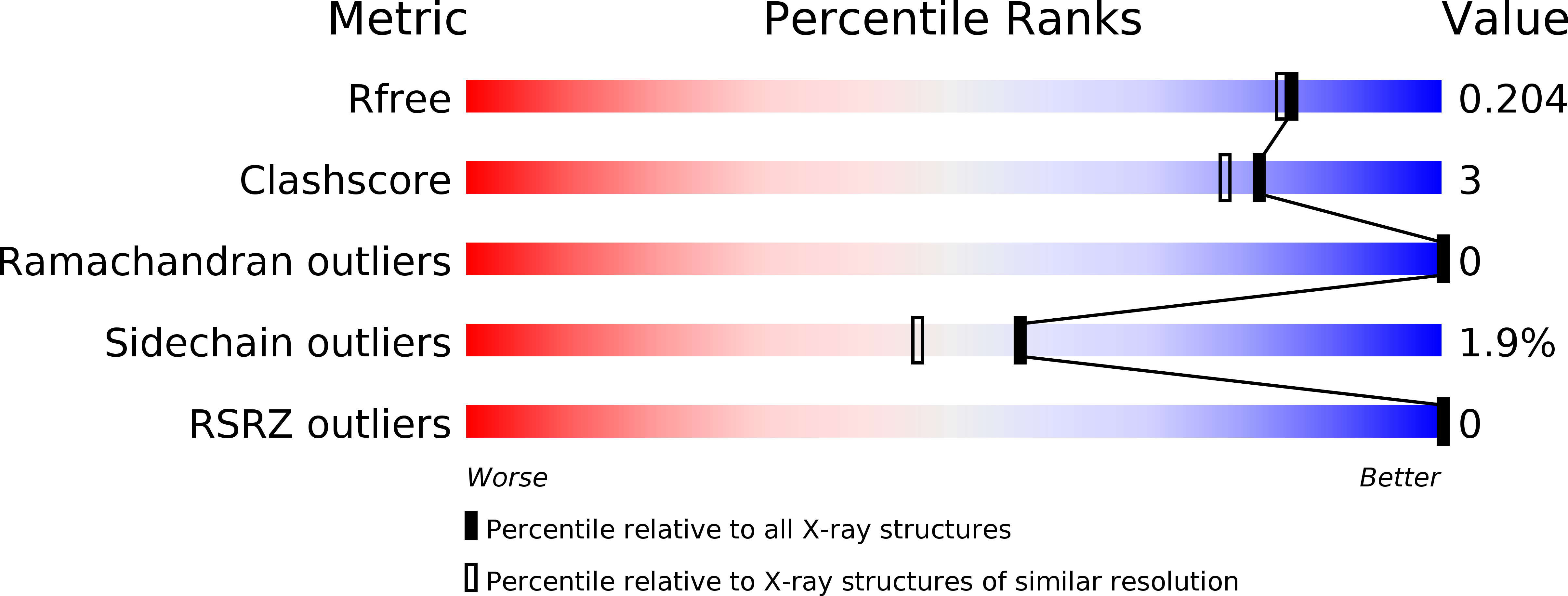
Resolution:
1.80 Å
R-Value Free:
0.19
R-Value Work:
0.16
R-Value Observed:
0.16
Space Group:
P 61 2 2
Macromolecular Entities
Polymer Type:polypeptide(L)
Molecule:UBA-domain protein mud1
Gene (Uniprot):mud1
Chain IDs:A, B
Chain Length:40
Number of Molecules:2
Biological Source:Schizosaccharomyces pombe
Primary Citation
Mechanism of Lys48-linked polyubiquitin chain recognition by the Mud1 UBA domain
Embo J. 24 3178 3189 (2005)
PMID: 16138082 DOI: 10.1038/sj.emboj.7600797

Abstact

The ubiquitin-pathway associated (UBA) domain is a 40-residue polyubiquitin-binding motif. The Schizosaccharomyces pombe protein Mud1 is an ortholog of the Saccharomyces cerevisiae DNA-damage response protein Ddi1 and binds to K48-linked polyubiquitin through its UBA domain. We have solved the crystal structure of Mud1 UBA at 1.8 angstroms resolution, revealing a canonical three-helical UBA fold. We have probed the interactions of this domain using mutagenesis, surface plasmon resonance, NMR and analytical ultracentrifugation. We show that the ubiquitin-binding surface of Mud1 UBA extends beyond previously recognized motifs and can be functionally dissected into primary and secondary ubiquitin-binding sites. Mutation of Phe330 to alanine, a residue exposed between helices 2 and 3, significantly reduces the affinity of the Mud1 UBA domain for K48-linked polyubiquitin, despite leaving the primary binding surface functionally intact. Moreover, K48-linked diubiquitin binds a single Mud1 UBA domain even in the presence of excess UBA. We therefore propose a mechanism for the recognition of K48-linked polyubiquitin chains by Mud1 in which diubiquitin units are specifically recognized by a single UBA domain.

Legend

Protein

Chemical

Disease

Primary Citation of related structures