1Z8Y image
Entry Detail
PDB ID:
1Z8Y
Keywords:
Title:
Mapping the E2 Glycoprotein of Alphaviruses
Biological Source:
Source Organism:
PDB Version:
Deposition Date:
2005-03-31
Release Date:
2006-02-07
Method Details:
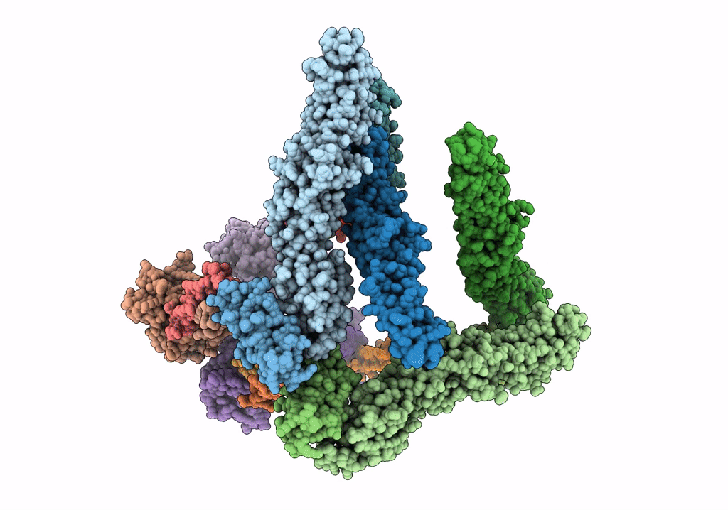
Experimental Method:
Resolution:
9.00 Å
Aggregation State:
PARTICLE
Reconstruction Method:
SINGLE PARTICLE
Macromolecular Entities
Polymer Type:polypeptide(L)
Description:Spike glycoprotein E1
Chain IDs:A, C, E, G
Chain Length:290
Number of Molecules:4
Biological Source:Sindbis virus
Polymer Type:polypeptide(L)
Description:Spike glycoprotein E1
Chain IDs:B, D, F, H
Chain Length:89
Number of Molecules:4
Biological Source:Sindbis virus
Polymer Type:polypeptide(L)
Description:Spike glycoprotein E1
Chain IDs:I, K, M, O
Chain Length:31
Number of Molecules:4
Biological Source:Sindbis virus
Polymer Type:polypeptide(L)
Description:Spike glycoprotein E2
Chain IDs:J, L, N, P
Chain Length:36
Number of Molecules:4
Biological Source:Sindbis virus
Polymer Type:polypeptide(L)
Description:Capsid protein C
Chain IDs:Q, R, S, T
Chain Length:151
Number of Molecules:4
Biological Source:Sindbis virus
Ligand Molecules
Primary Citation
Mapping the structure and function of the E1 and E2 glycoproteins in alphaviruses.
Structure 14 63 73 (2006)
PMID: 16407066 DOI: 10.1016/j.str.2005.07.025

Abstact

The 9 A resolution cryo-electron microscopy map of Sindbis virus presented here provides structural information on the polypeptide topology of the E2 protein, on the interactions between the E1 and E2 glycoproteins in the formation of a heterodimer, on the difference in conformation of the two types of trimeric spikes, on the interaction between the transmembrane helices of the E1 and E2 proteins, and on the conformational changes that occur when fusing with a host cell. The positions of various markers on the E2 protein established the approximate topology of the E2 structure. The largest conformational differences between the icosahedral surface spikes at icosahedral 3-fold and quasi-3-fold positions are associated with the monomers closest to the 5-fold axes. The long E2 monomers, containing the cell receptor recognition motif at their extremities, are shown to rotate by about 180 degrees and to move away from the center of the spikes during fusion.

Legend

Protein

Chemical

Disease

Primary Citation of related structures