
Deposition Date
2005-03-26
Release Date
2005-08-09
Last Version Date
2023-08-23
Entry Detail
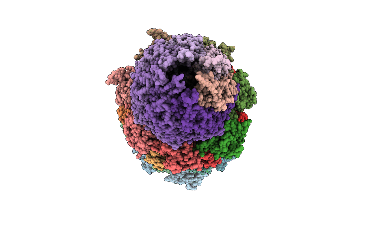
PDB ID:
1Z7Q
Keywords:
Title:
Crystal structure of the 20s proteasome from yeast in complex with the proteasome activator PA26 from Trypanosome brucei at 3.2 angstroms resolution
Biological Source:
Source Organism(s):
Saccharomyces cerevisiae (Taxon ID: 4932)
Trypanosoma brucei (Taxon ID: 5691)
Trypanosoma brucei (Taxon ID: 5691)
Expression System(s):
Method Details:
Experimental Method:
Resolution:
3.22 Å
R-Value Free:
0.30
R-Value Work:
0.26
R-Value Observed:
0.26
Space Group:
P 21 21 21
