1Z7K image
Deposition Date 2005-03-25
Release Date 2005-04-05
Last Version Date 2024-11-13
Entry Detail
PDB ID:
1Z7K
Title:
Crystal Structure of Trypsin- Ovomucoid turkey egg white inhibitor complex
Biological Source:
Source Organism:
Sus scrofa (Taxon ID: 9823)
Meleagris gallopavo (Taxon ID: 9103)
Method Details:
Experimental Method:
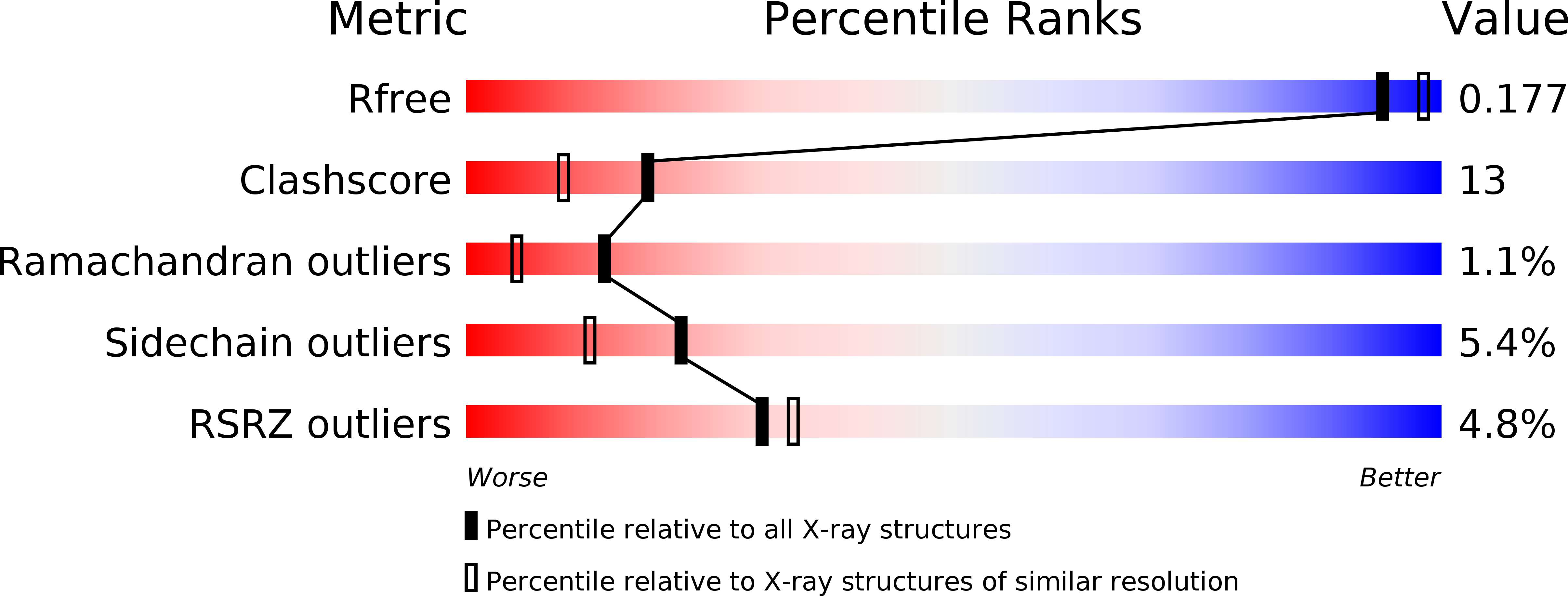
Resolution:
1.90 Å
R-Value Free:
0.18
R-Value Work:
0.17
R-Value Observed:
0.17
Space Group:
P 65
Macromolecular Entities
Polymer Type:polypeptide(L)
Molecule:Trypsin
Chain IDs:A
Chain Length:223
Number of Molecules:1
Biological Source:Sus scrofa
Polymer Type:polypeptide(L)
Molecule:Ovomucoid
Chain IDs:B
Chain Length:62
Number of Molecules:1
Biological Source:Meleagris gallopavo
Polymer Type:polypeptide(L)
Molecule:Ovomucoid
Chain IDs:C
Chain Length:4
Number of Molecules:1
Biological Source:Meleagris gallopavo
Modified Residue
Compound ID Chain ID Parent Comp ID Details 2D Image
ASN B ASN GLYCOSYLATION SITE
Primary Citation
Crystal structure of trypsin-turkey egg white inhibitor complex
Biochem.Biophys.Res.Commun. 313 8 16 (2004)
PMID: 14672690 DOI: 10.1016/j.bbrc.2003.11.082

Abstact

Crystal structure of the complex between porcine beta-trypsin and the second domain of the Kazal-type ovomucoid turkey egg white trypsin inhibitor (OMTKY2) has been determined at 1.9A resolution. A peptide fragment from the first domain has been crystallized with the complex. Restrained-refinement of the structure led to an R-factor of 0.19 for the 32206 reflections. OMTKY2 exhibits the canonical Kazal-type fold with a central alpha-helix and a short two-stranded anti-parallel beta-sheet. The carbonyl carbon of the reactive site prefers trigonal geometry. The reactive site loop geometry of the inhibitor is complementary to the surface and charge of the binding site in beta-trypsin.

Legend

Protein

Chemical

Disease

Primary Citation of related structures