Deposition Date
2005-03-22
Release Date
2005-06-21
Last Version Date
2024-11-20
Entry Detail
PDB ID:
1Z6F
Keywords:
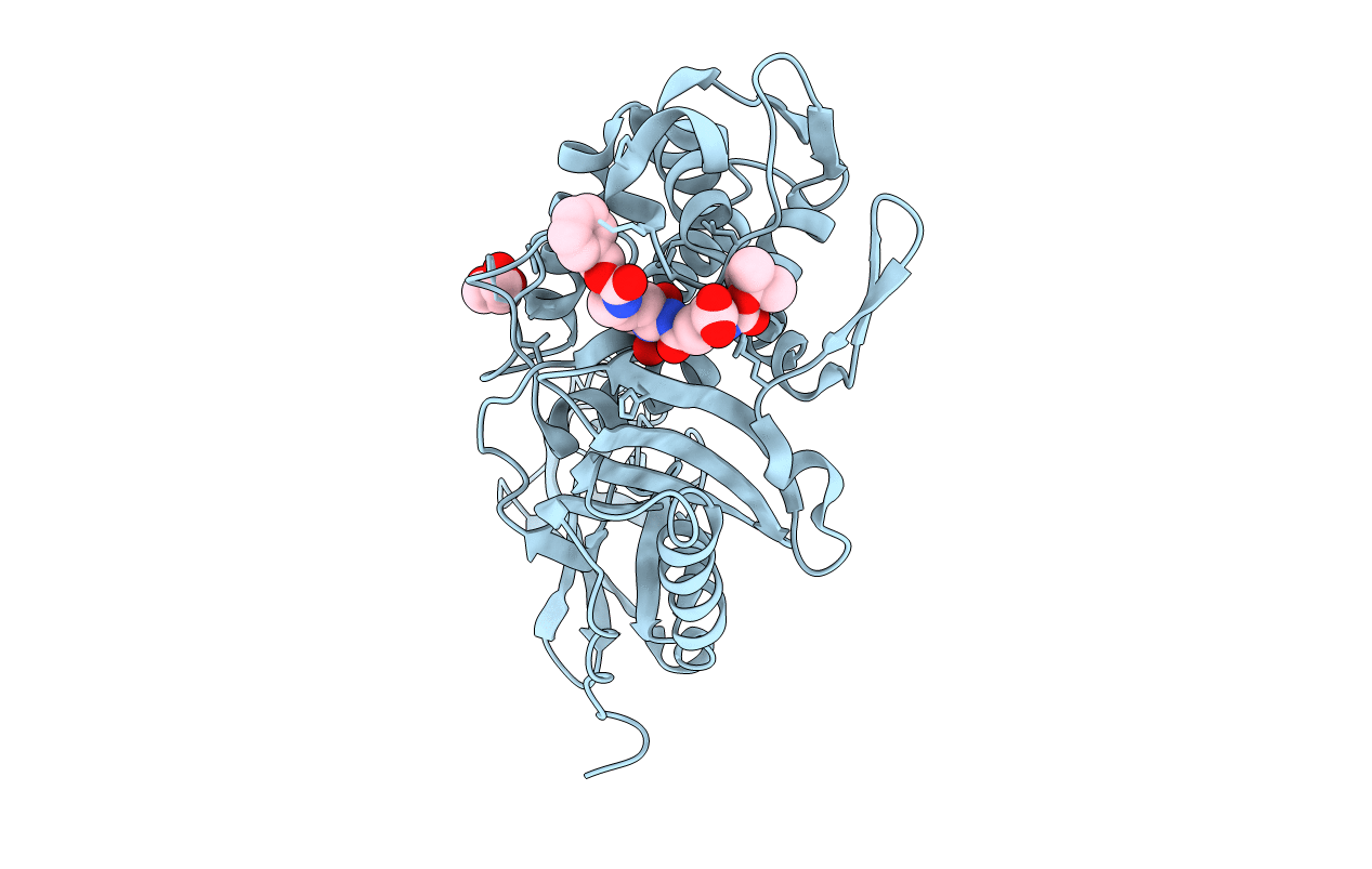
Title:
Crystal structure of penicillin-binding protein 5 from E. coli in complex with a boronic acid inhibitor
Biological Source:
Source Organism(s):
Escherichia coli (Taxon ID: 562)
Expression System(s):
Method Details:
Experimental Method:
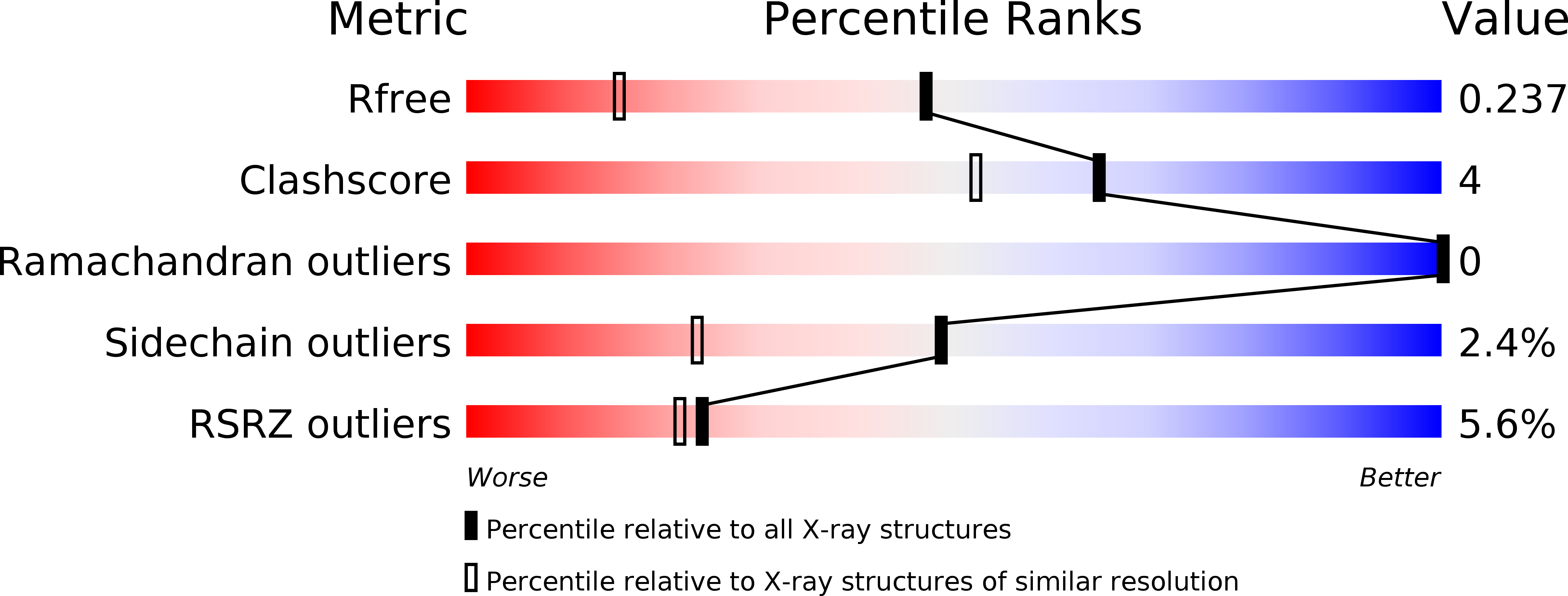
Resolution:
1.60 Å
R-Value Free:
0.24
R-Value Work:
0.21
R-Value Observed:
0.21
Space Group:
C 1 2 1