1Z3U image
Deposition Date 2005-03-14
Release Date 2005-07-12
Last Version Date 2024-10-30
Entry Detail
PDB ID:
1Z3U
Title:
Structure of the Angiopoietin-2 Recptor Binding Domain and Identification of Surfaces Involved in Tie2 Recognition
Biological Source:
Source Organism:
Homo sapiens (Taxon ID: 9606)
Host Organism:
Method Details:
Experimental Method:
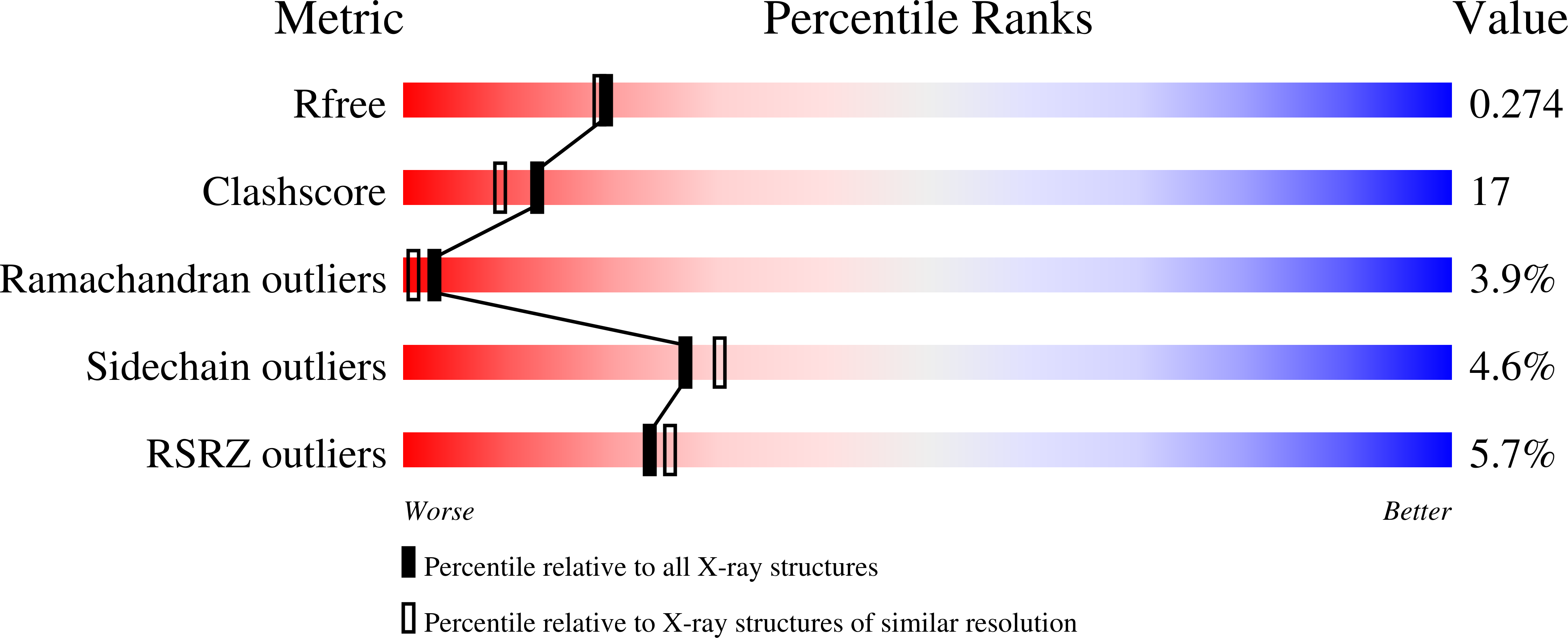
Resolution:
2.25 Å
R-Value Free:
0.27
R-Value Work:
0.24
R-Value Observed:
0.24
Space Group:
C 1 2 1
Macromolecular Entities
Polymer Type:polypeptide(L)
Molecule:Angiopoietin-2
Gene (Uniprot):ANGPT2
Mutations:F469A, Y475A, Y476A
Chain IDs:A, B, C, D
Chain Length:217
Number of Molecules:4
Biological Source:Homo sapiens
Ligand Molecules
Primary Citation
Structure of the angiopoietin-2 receptor binding domain and identification of surfaces involved in Tie2 recognition.
Structure 13 825 832 (2005)
PMID: 15893672 DOI: 10.1016/j.str.2005.03.009

Abstact

The angiopoietins comprise a small class of secreted glycoproteins that play crucial roles in the maturation and maintenance of the mammalian vascular and lymphatic systems. They exert their effects through a member of the tyrosine kinase receptor family, Tie2. Angiopoietin/Tie2 signaling is unique among tyrosine kinase receptor-ligand systems in that distinct angiopoietin ligands, although highly homologous, can function as agonists or antagonists in a context-dependent manner. In an effort to understand this molecular dichotomy, we have crystallized and determined the 2.4 A crystal structure of the Angiopoietin-2 (Ang2) receptor binding region. The structure reveals a fibrinogen fold with a unique C-terminal P domain. Conservation analysis and structure-based mutagenesis identify a groove on the Ang2 molecular surface that mediates receptor recognition.

Legend

Protein

Chemical

Disease

Primary Citation of related structures