Deposition Date
2005-03-08
Release Date
2005-05-24
Last Version Date
2024-02-14
Entry Detail
PDB ID:
1Z2M
Keywords:
Title:
Crystal Structure of ISG15, the Interferon-Induced Ubiquitin Cross Reactive Protein
Biological Source:
Source Organism(s):
Homo sapiens (Taxon ID: 9606)
Expression System(s):
Method Details:
Experimental Method:
Resolution:
2.50 Å
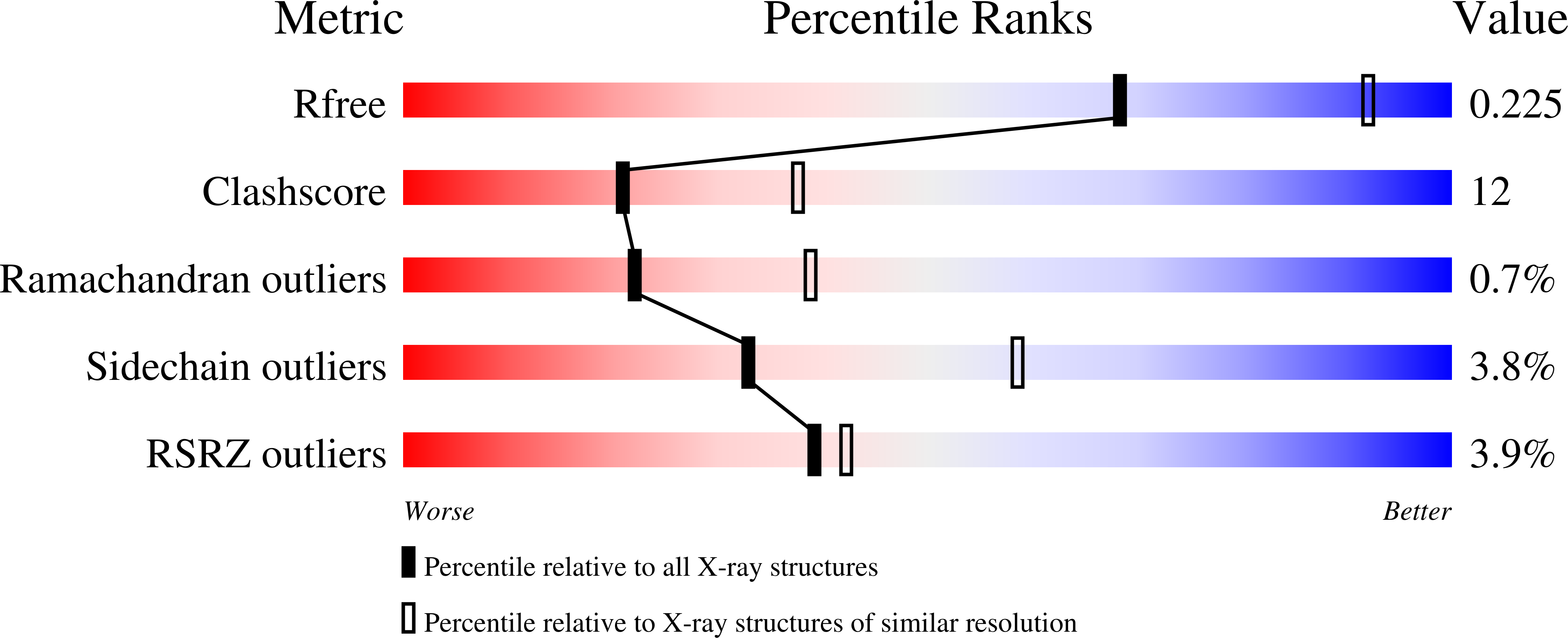
R-Value Free:
0.22
R-Value Work:
0.20
R-Value Observed:
0.20
Space Group:
I 2 2 2