1Z2C image
Deposition Date 2005-03-08
Release Date 2005-05-10
Last Version Date 2024-02-14
Entry Detail
PDB ID:
1Z2C
Title:
Crystal structure of mDIA1 GBD-FH3 in complex with RhoC-GMPPNP
Biological Source:
Source Organism:
Homo sapiens (Taxon ID: 9606)
Mus musculus (Taxon ID: 10090)
Method Details:
Experimental Method:
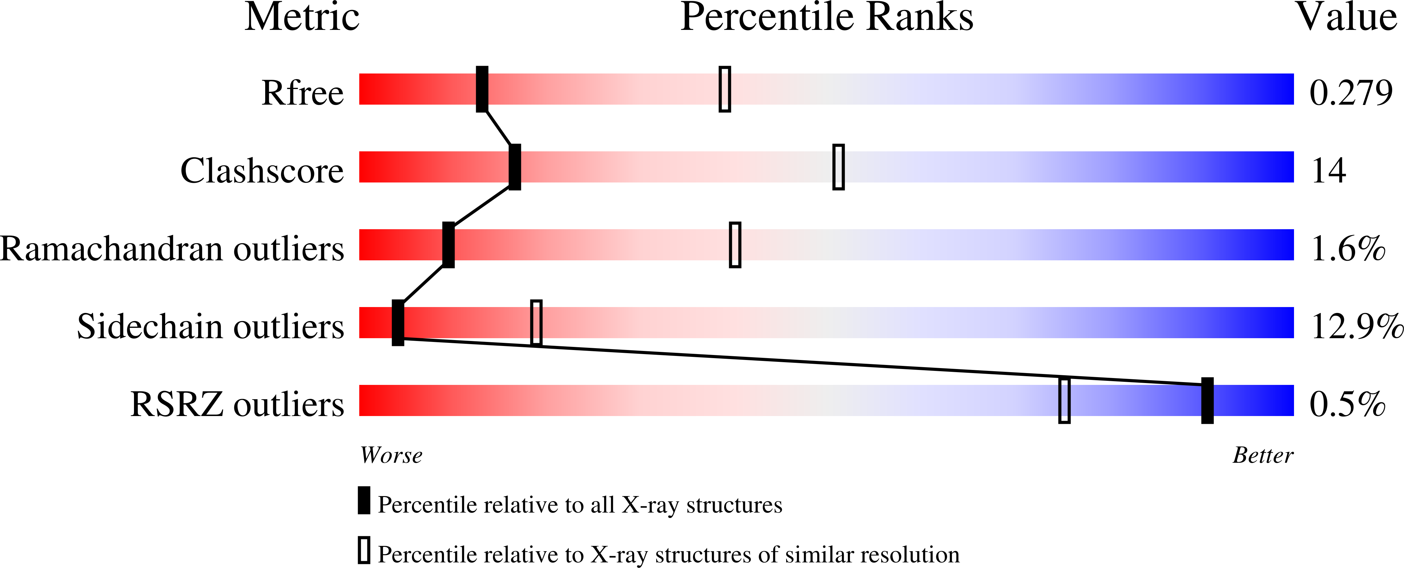
Resolution:
3.00 Å
R-Value Free:
0.28
R-Value Work:
0.21
R-Value Observed:
0.21
Space Group:
P 21 21 2
Macromolecular Entities
Polymer Type:polypeptide(L)
Molecule:Rho-related GTP-binding protein RhoC
Gene (Uniprot):RHOC
Mutations:F25N
Chain IDs:A, C
Chain Length:193
Number of Molecules:2
Biological Source:Homo sapiens
Polymer Type:polypeptide(L)
Molecule:Diaphanous protein homolog 1
Gene (Uniprot):Diaph1
Chain IDs:B, D
Chain Length:383
Number of Molecules:2
Biological Source:Mus musculus
Primary Citation
Structural and mechanistic insights into the interaction between Rho and mammalian Dia.
Nature 435 513 518 (2005)
PMID: 15864301 DOI: 10.1038/nature03604

Abstact

Formins are involved in a variety of cellular processes that require the remodelling of the cytoskeleton. They contain formin homology domains FH1 and FH2, which initiate actin assembly. The Diaphanous-related formins form a subgroup that is characterized by an amino-terminal Rho GTPase-binding domain (GBD) and an FH3 domain, which bind somehow to the carboxy-terminal Diaphanous autoregulatory domain (DAD) to keep the protein in an inactive conformation. Upon binding of activated Rho proteins, the DAD is released and the ability of the formin to nucleate and elongate unbranched actin filaments is induced. Here we present the crystal structure of RhoC in complex with the regulatory N terminus of mammalian Diaphanous 1 (mDia1) containing the GBD/FH3 region, an all-helical structure with armadillo repeats. Rho uses its 'switch' regions for interacting with two subdomains of GBD/FH3. We show that the FH3 domain of mDia1 forms a stable dimer and we also identify the DAD-binding site. Although binding of Rho and DAD on the N-terminal fragment of mDia1 are mutually exclusive, their binding sites are only partially overlapping. On the basis of our results, we propose a structural model for the regulation of mDia1 by Rho and DAD.

Legend

Protein

Chemical

Disease

Primary Citation of related structures