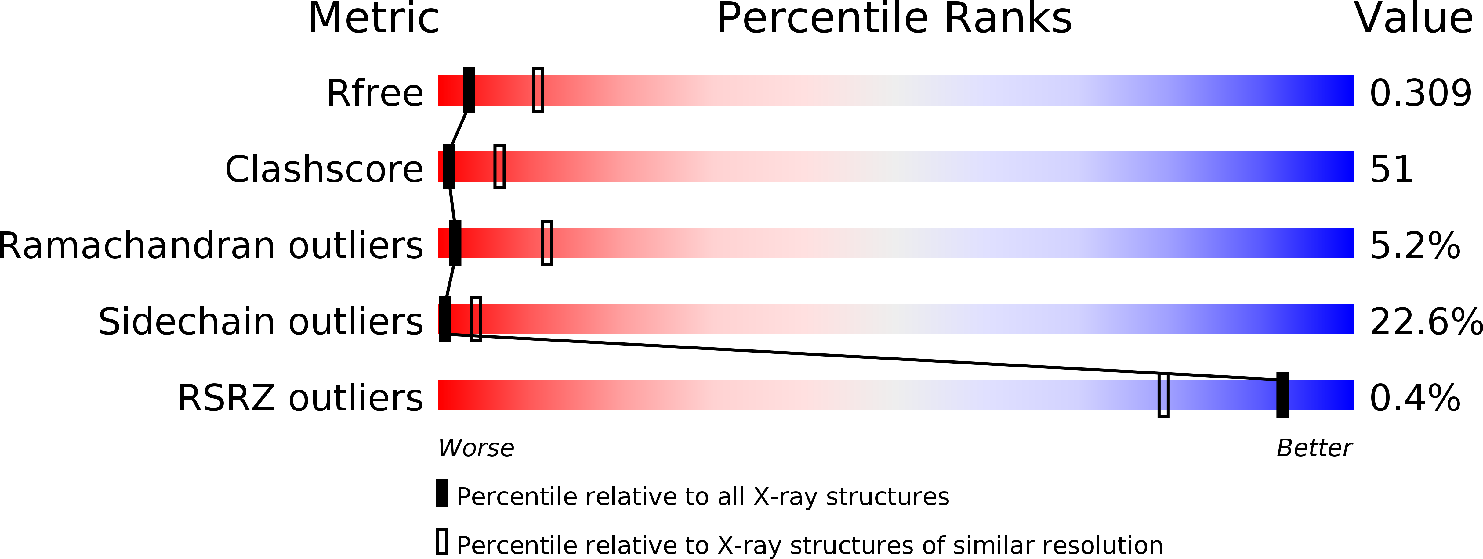
1Z0V image
Deposition Date 2005-03-02
Release Date 2005-08-02
Last Version Date 2024-02-14
Entry Detail
PDB ID:
1Z0V
Keywords:
Title:
Crystal Structure of A. fulgidus Lon proteolytic domain
Biological Source:
Source Organism:
Host Organism:
Method Details:
Experimental Method:
Resolution:
3.00 Å
R-Value Free:
0.31
R-Value Work:
0.21
R-Value Observed:
0.21
Space Group:
P 1 21 1
Macromolecular Entities
Polymer Type:polypeptide(L)
Molecule:Putative protease La homolog type
Gene (Uniprot):AF_0364
Chain IDs:A, B, C, D, E, F
Chain Length:205
Number of Molecules:6
Biological Source:Archaeoglobus fulgidus
Primary Citation
Pathological crystallography: case studies of several unusual macromolecular crystals.
Acta Crystallogr.,Sect.D 61 967 975 (2005)
PMID: 15983420 DOI: 10.1107/S0907444905011285

Abstact

Although macromolecular crystallography is rapidly becoming largely routine owing to advances in methods of data collection, structure solution and refinement, difficult cases are still common. To remind structural biologists about the kinds of crystallographic difficulties that might be encountered, case studies of several successfully completed structure determinations that utilized less than perfect crystals are discussed here. The structure of the proteolytic domain of Archaeoglobus fulgidus Lon was solved with crystals that contained superimposed orthorhombic and monoclinic lattices, a case not previously described for proteins. Another hexagonal crystal form of this protein exhibited an unusually high degree of non-isomorphism. Crystals of A. fulgidus Rio1 kinase exhibited both pseudosymmetry and twinning. Ways of identifying the observed phenomena and approaches to solving and refining macromolecular structures when only less than perfect crystals are available are discussed here.

Legend

Protein

Chemical

Disease

Primary Citation of related structures