
Deposition Date
2005-03-02
Release Date
2005-10-25
Last Version Date
2024-10-23
Entry Detail
PDB ID:
1Z0N
Keywords:
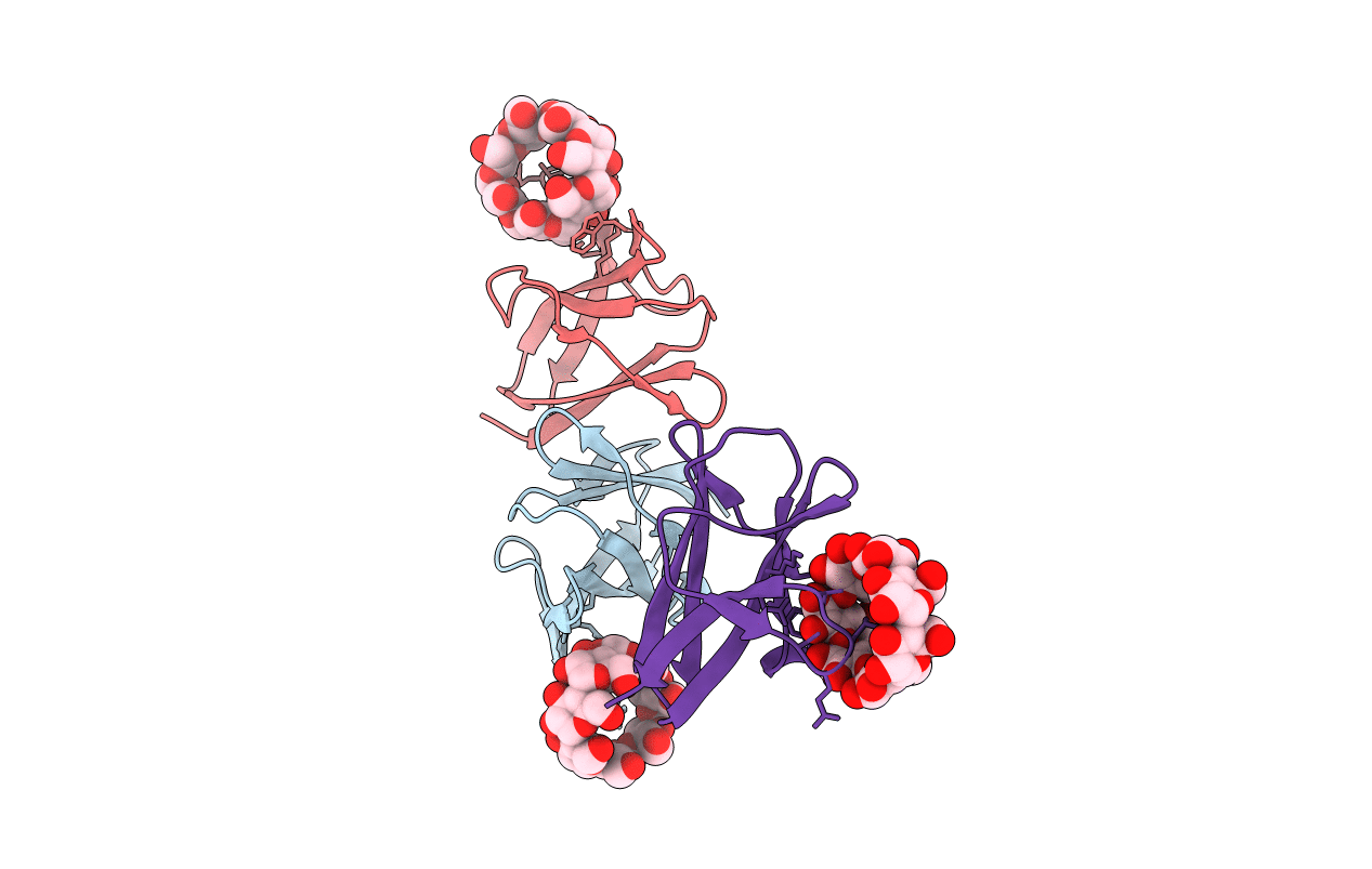
Title:
the glycogen-binding domain of the AMP-activated protein kinase
Biological Source:
Source Organism(s):
Rattus norvegicus (Taxon ID: 10116)
Expression System(s):
Method Details:
Experimental Method:
Resolution:
1.49 Å
R-Value Free:
0.21
R-Value Work:
0.18
R-Value Observed:
0.18
Space Group:
P 1
