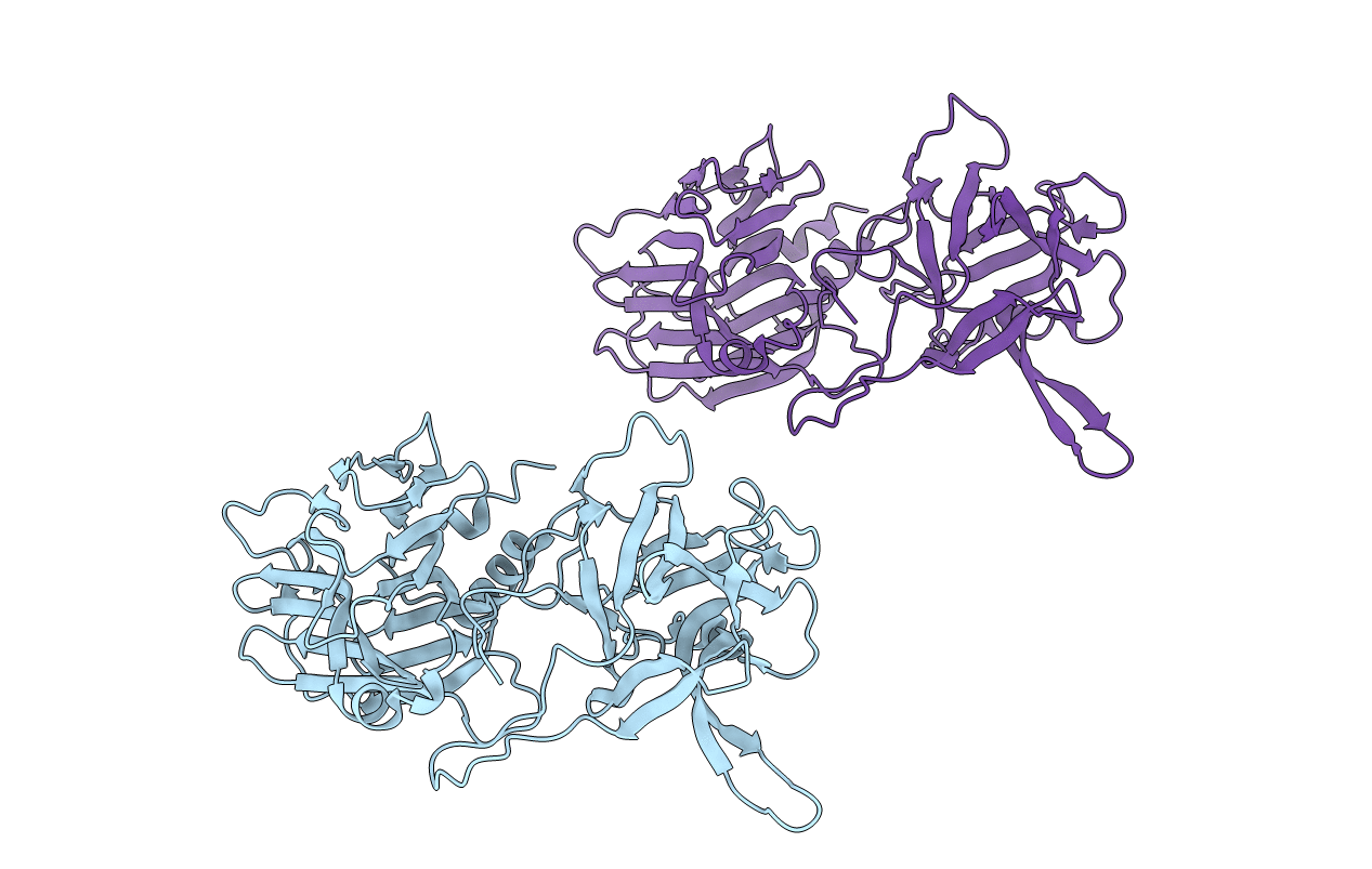
1Z0H image
Deposition Date 2005-03-01
Release Date 2005-03-15
Last Version Date 2023-08-23
Entry Detail
PDB ID:
1Z0H
Keywords:
Title:
N-terminal helix reorients in recombinant C-fragment of Clostridium botulinum type B
Biological Source:
Source Organism:
Host Organism:
Method Details:
Experimental Method:
Resolution:
2.00 Å
R-Value Free:
0.28
R-Value Work:
0.23
R-Value Observed:
0.23
Space Group:
P 1 21 1
Macromolecular Entities
Structures with similar UniProt ID
Protein Blast
Polymer Type:polypeptide(L)
Molecule:Botulinum neurotoxin type B
Gene (Uniprot):botB
Chain IDs:A, B
Chain Length:438
Number of Molecules:2
Biological Source:Clostridium botulinum
Primary Citation
N-terminal helix reorients in recombinant C-fragment of Clostridium botulinum type B.
Biochem.Biophys.Res.Commun. 330 97 103 (2005)
PMID: 15781237 DOI: 10.1016/j.bbrc.2005.02.123

Abstact

Botulinum neurotoxins comprise seven distinct serotypes (A-G) produced by Clostridium botulinum. The crystal structure of the binding domain of the botulinum neurotoxin type B (BBHc) has been determined to 2A resolution. The overall structure of BBHc is well ordered and similar to that of the binding domain of the holotoxin. However, significant structural changes occur at what would be the interface of translocation and binding domains of the holotoxin. The loop 911-924 shows a maximum displacement of 14.8A at the farthest point. The N-terminal helix reorients and moves by 19.5A from its original position. BBHc is compared with the binding domain of the holotoxin of botulinum type A and B, and the tetanus C-fragment to characterize the heavy chain-carbohydrate interactions. The probable reasons for different binding affinity of botulinum and tetanus toxins are discussed.

Legend

Protein

Chemical

Disease

Primary Citation of related structures