1YZI image
Deposition Date 2005-02-28
Release Date 2005-03-15
Last Version Date 2023-08-23
Entry Detail
PDB ID:
1YZI
Title:
A novel quaternary structure of human carbonmonoxy hemoglobin
Biological Source:
Source Organism:
Homo sapiens (Taxon ID: 9606)
Method Details:
Experimental Method:
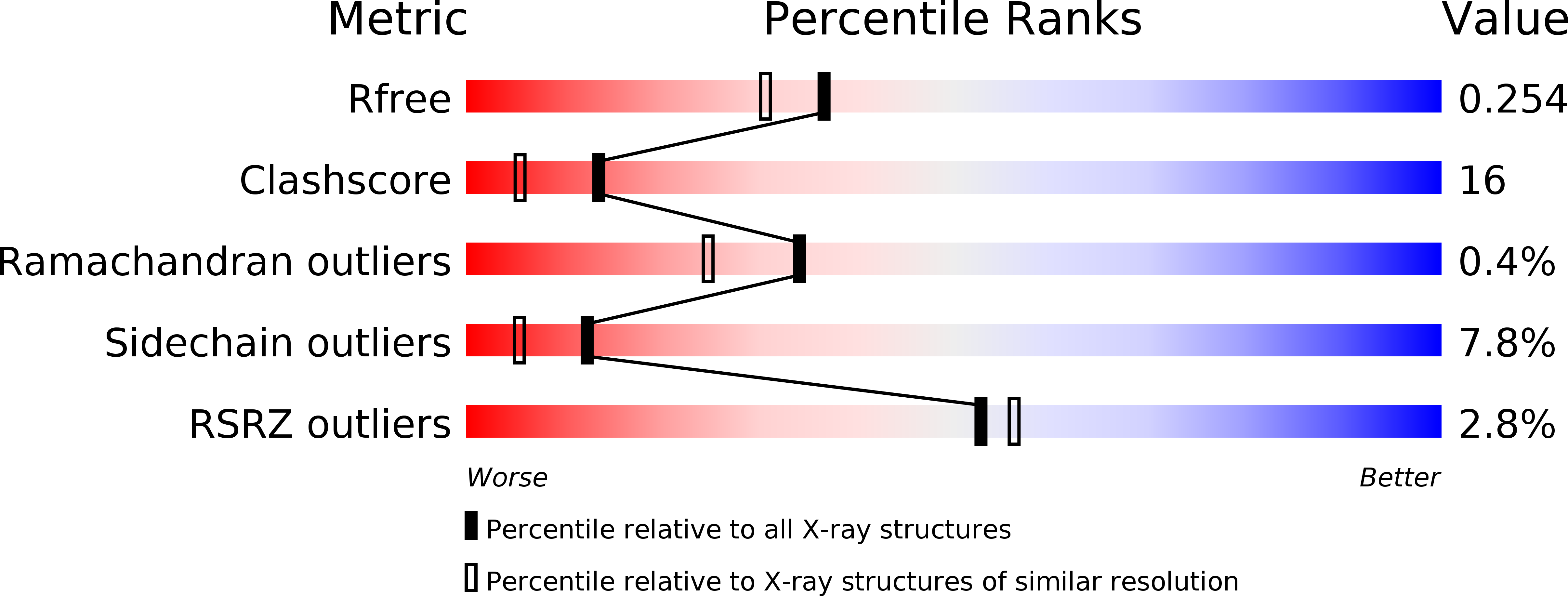
Resolution:
2.07 Å
R-Value Free:
0.25
R-Value Work:
0.20
R-Value Observed:
0.20
Space Group:
P 41 2 2
Macromolecular Entities
Structures with similar UniProt ID
Protein Blast
Polymer Type:polypeptide(L)
Molecule:Hemoglobin alpha chain
Gene (Uniprot):HBA1, HBA2
Chain IDs:A
Chain Length:141
Number of Molecules:1
Biological Source:Homo sapiens
Structures with similar UniProt ID
Protein Blast
Polymer Type:polypeptide(L)
Molecule:Hemoglobin beta chain
Gene (Uniprot):HBB
Chain IDs:B
Chain Length:146
Number of Molecules:1
Biological Source:Homo sapiens
Primary Citation
The enigma of the liganded hemoglobin end state: a novel quaternary structure of human carbonmonoxy hemoglobin.
Biochemistry 44 8347 8359 (2005)
PMID: 15938624 DOI: 10.1021/bi050412q

Abstact

The liganded hemoglobin (Hb) high-salt crystallization condition described by Max Perutz has generated three different crystals of human adult carbonmonoxy hemoglobin (COHbA). The first crystal is isomorphous with the "classical" liganded or R Hb structure. The second crystal reveals a new liganded Hb quaternary structure, RR2, that assumes an intermediate conformation between the R form and another liganded Hb quaternary structure, R2, which was discovered more than a decade ago. Like the R2 structure, the diagnostic R state hydrogen bond between beta2His97 and alpha1Thr38 is missing in the RR2 structure. The third crystal adopts a novel liganded Hb conformation, which we have termed R3, and it shows substantial quaternary structural differences from the R, RR2, and R2 structures. The quaternary structure differences between T and R3 are as large as those between T and R2; however, the T --> R3 and T --> R2 transitions are in different directions as defined by rigid-body screw rotation. Moreover, R3 represents an end state. Compared to all known liganded Hb structures, R3 shows remarkably reduced strain at the alpha-heme, reduced steric contact between the beta-heme ligand and the distal residues, smaller alpha- and beta-clefts, and reduced alpha1-alpha2 and beta1-beta2 iron-iron distances. Together, these unique structural features in R3 should make it the most relaxed and/or greatly enhance its affinity for oxygen compared to the other liganded Hbs. The current Hb structure-function relationships that are now based on T --> R, T -->R --> R2, or T --> R2 --> R transitions may have to be reexamined to take into account the RR2 and R3 liganded structures.

Legend

Protein

Chemical

Disease

Primary Citation of related structures