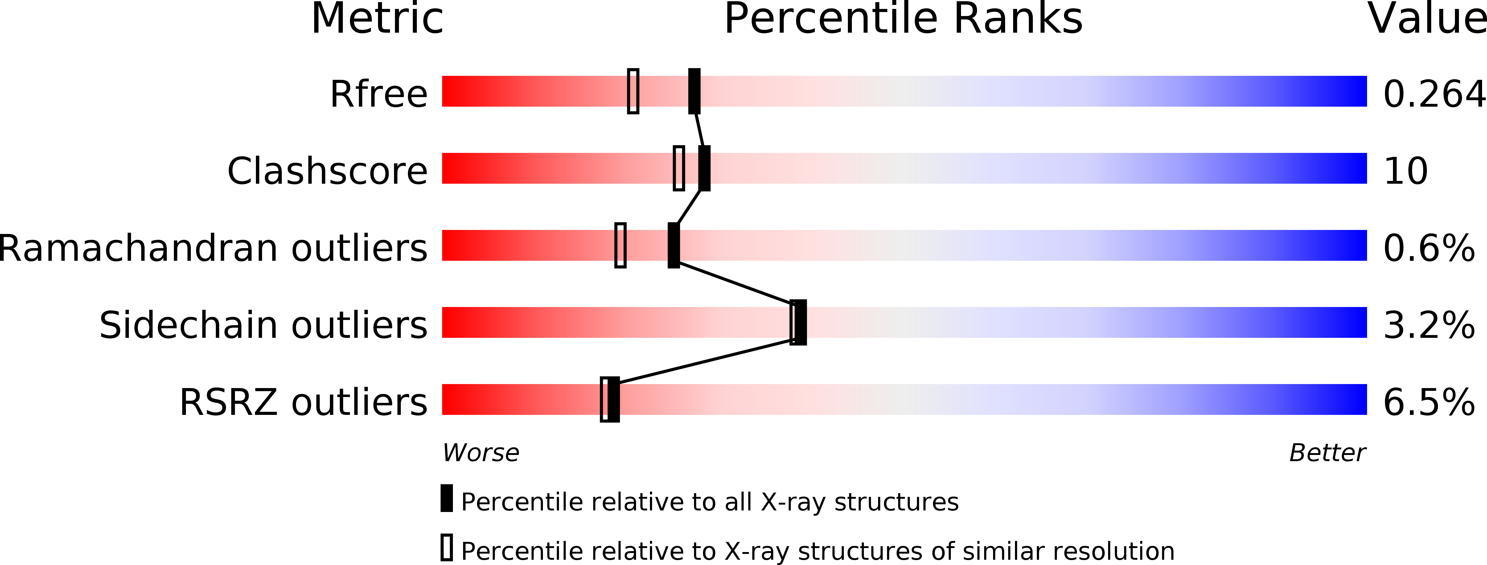
1YY8 image
Deposition Date 2005-02-24
Release Date 2005-04-26
Last Version Date 2024-11-20
Entry Detail
PDB ID:
1YY8
Keywords:
Title:
Crystal structure of the Fab fragment from the monoclonal antibody cetuximab/Erbitux/IMC-C225
Biological Source:
Source Organism:
Host Organism:
Method Details:
Experimental Method:
Resolution:
2.00 Å
R-Value Free:
0.26
R-Value Work:
0.22
Space Group:
P 21 21 21
Macromolecular Entities
Polymer Type:polypeptide(L)
Molecule:Cetuximab Fab Light chain
Chain IDs:A, C
Chain Length:213
Number of Molecules:2
Biological Source:Mus musculus, Homo sapiens
Polymer Type:polypeptide(L)
Molecule:Cetuximab Fab Heavy chain
Chain IDs:B, D
Chain Length:221
Number of Molecules:2
Biological Source:Mus musculus, Homo sapiens
Primary Citation
Structural basis for inhibition of the epidermal growth factor receptor by cetuximab
Cancer Cell 7 301 311 (2005)
PMID: 15837620 DOI: 10.1016/j.ccr.2005.03.003

Abstact

Recent structural studies of epidermal growth factor receptor (EGFR) family extracellular regions have identified an unexpected mechanism for ligand-induced receptor dimerization that has important implications for activation and inhibition of these receptors. Here we describe the 2.8 angstroms resolution X-ray crystal structure of the antigen binding (Fab) fragment from cetuximab (Erbitux), an inhibitory anti-EGFR antibody, in complex with the soluble extracellular region of EGFR (sEGFR). The sEGFR is in the characteristic "autoinhibited" or "tethered" inactive configuration. Cetuximab interacts exclusively with domain III of sEGFR, partially occluding the ligand binding region on this domain and sterically preventing the receptor from adopting the extended conformation required for dimerization. We suggest that both these effects contribute to potent inhibition of EGFR activation.

Legend

Protein

Chemical

Disease

Primary Citation of related structures