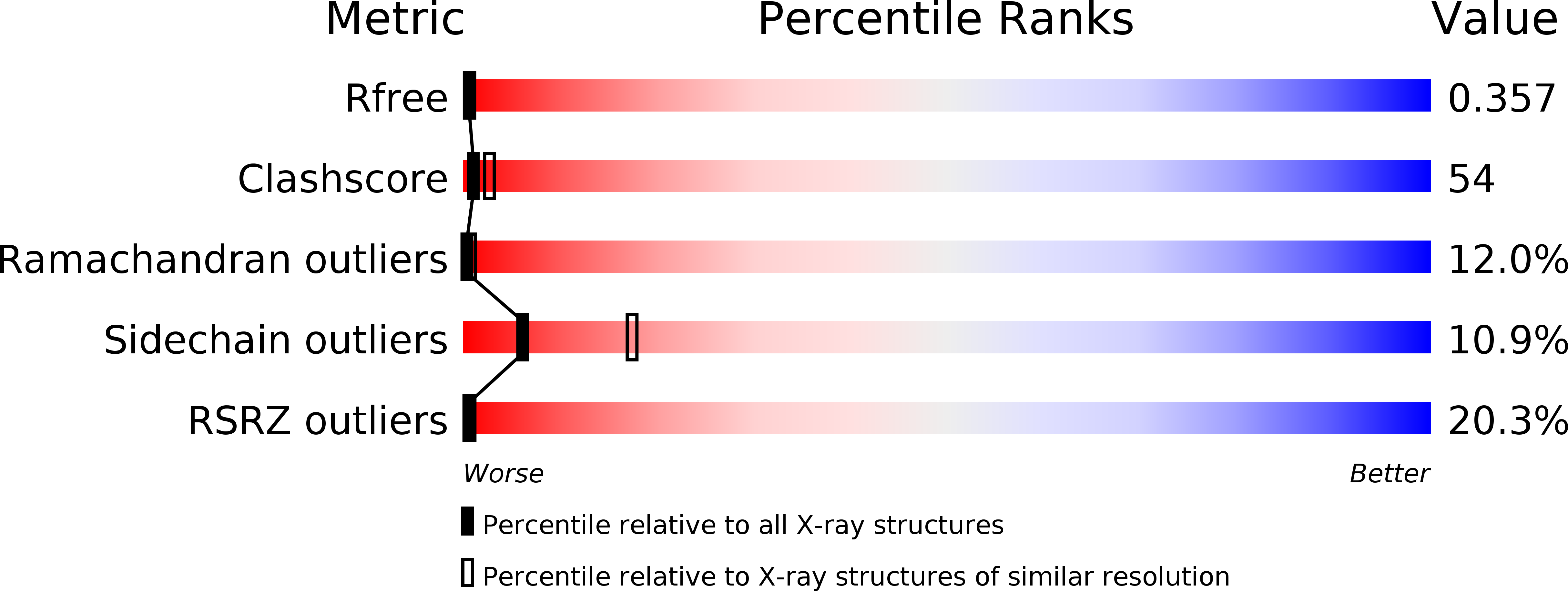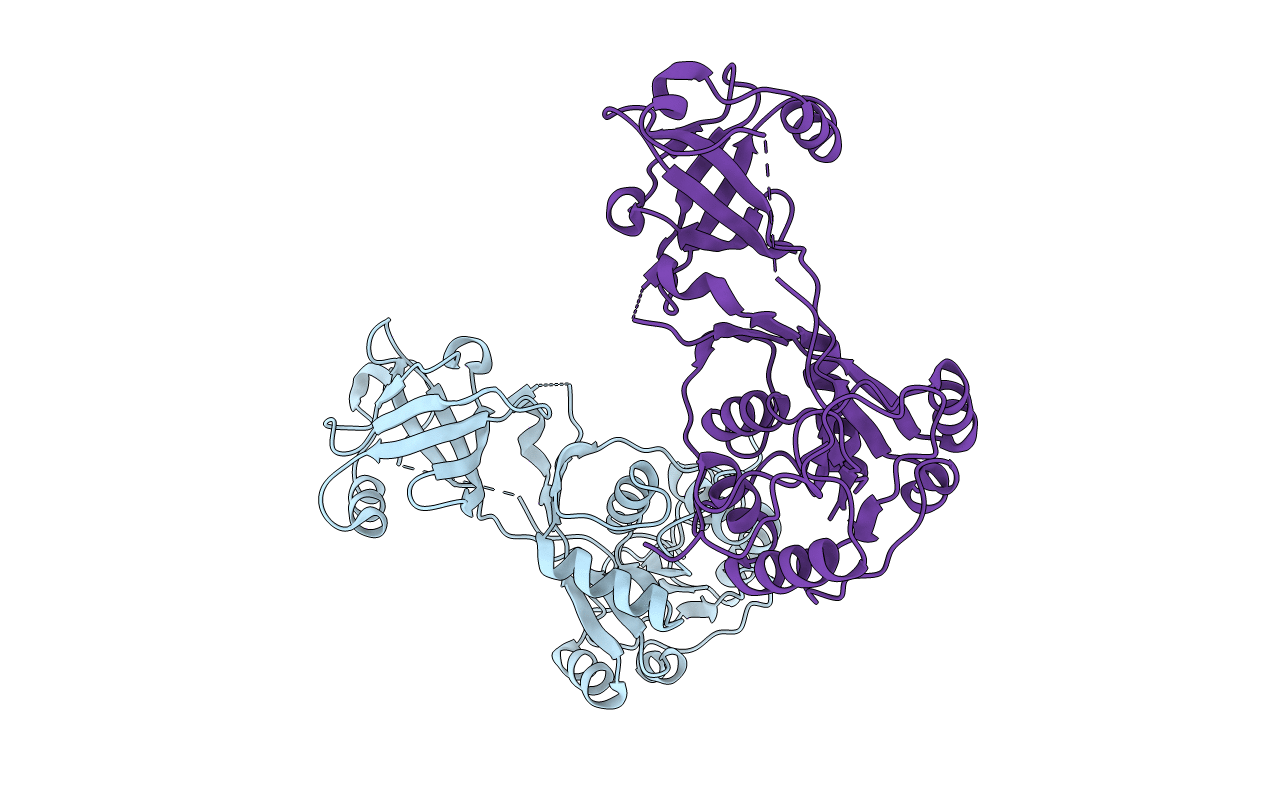
1YY3 image
Deposition Date 2005-02-23
Release Date 2006-03-21
Last Version Date 2024-03-13
Entry Detail
PDB ID:
1YY3
Keywords:
Title:
Structure of S-Adenosylmethionine:tRNA Ribosyltransferase-Isomerase (QueA)
Biological Source:
Source Organism:
Host Organism:
Method Details:
Experimental Method:
Resolution:
2.88 Å
R-Value Free:
0.38
R-Value Work:
0.36
R-Value Observed:
0.36
Space Group:
P 4 21 2
Macromolecular Entities
Structures with similar UniProt ID
Protein Blast
Polymer Type:polypeptide(L)
Molecule:S-adenosylmethionine:tRNA ribosyltransferase-isomerase
Gene (Uniprot):queA
Chain IDs:A, B
Chain Length:346
Number of Molecules:2
Biological Source:Bacillus subtilis
Ligand Molecules
Primary Citation
Crystal structure of Bacillus subtilis S-adenosylmethionine:tRNA ribosyltransferase-isomerase
Biochem.Biophys.Res.Commun. 351 695 701 (2006)
PMID: 17083917 DOI: 10.1016/j.bbrc.2006.10.096

Abstact

The enzyme S-adenosylmethionine:tRNA ribosyltransferase-isomerase (QueA) is involved in the biosynthesis of the hypermodified tRNA nucleoside queuosine. It is unprecedented in nature as it uses the cofactor S-adenosylmethionine as the donor of a ribosyl group. We have determined the crystal structure of Bacillus subtilis QueA at a resolution of 2.9A. The structure reveals two domains representing a 6-stranded beta-barrel and an alpha beta alpha-sandwich, respectively. All amino acid residues invariant in the QueA enzymes of known sequence cluster at the interface of the two domains indicating the localization of the substrate binding region and active center. Comparison of the B. subtilis QueA structure with the structure of QueA from Thermotoga maritima suggests a high domain flexibility of this enzyme.

Legend

Protein

Chemical

Disease

Primary Citation of related structures