1YXQ image
Deposition Date 2005-02-22
Release Date 2005-05-17
Last Version Date 2025-03-26
Entry Detail
PDB ID:
1YXQ
Title:
Crystal structure of actin in complex with swinholide A
Biological Source:
Source Organism:
Method Details:
Experimental Method:
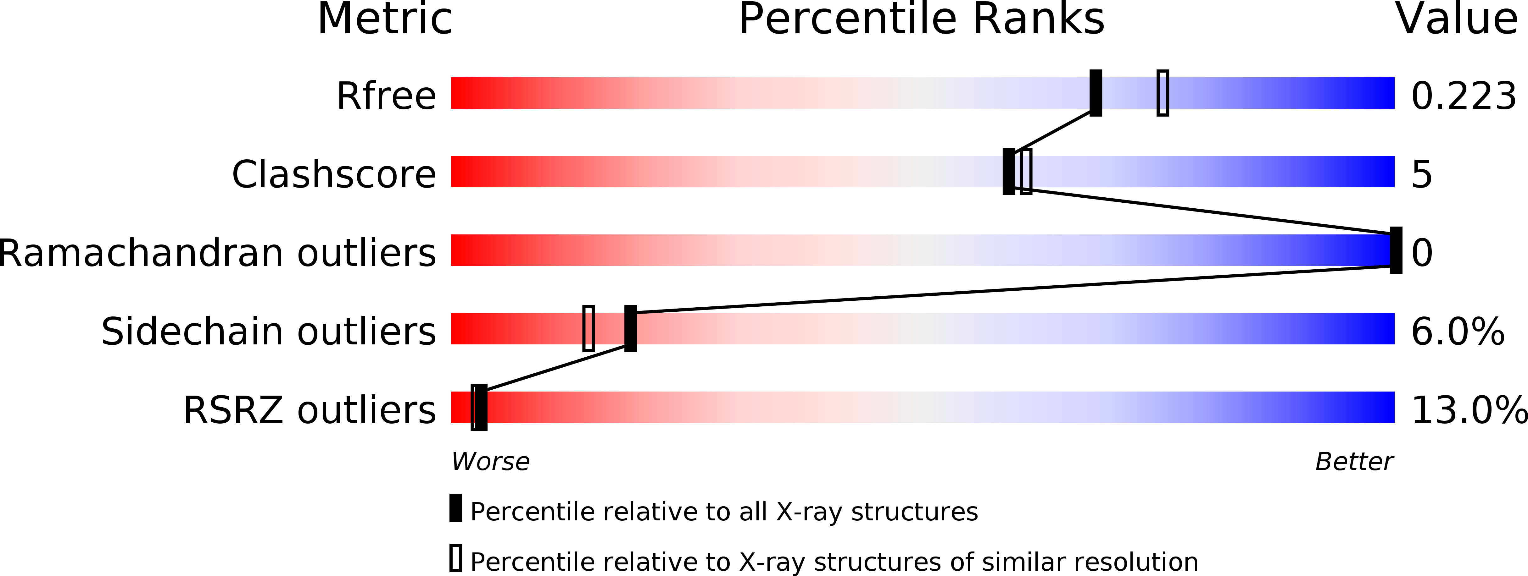
Resolution:
2.01 Å
R-Value Free:
0.21
R-Value Work:
0.18
R-Value Observed:
0.18
Space Group:
P 1 21 1
Macromolecular Entities
Polymer Type:polypeptide(L)
Molecule:actin, alpha skeletal muscle
Gene (Uniprot):ACTA1
Chain IDs:A, B
Chain Length:375
Number of Molecules:2
Biological Source:Oryctolagus cuniculus
Modified Residue
Compound ID Chain ID Parent Comp ID Details 2D Image
HIC A HIS 4-METHYL-HISTIDINE
Primary Citation
Structural basis of swinholide a binding to actin
Chem.Biol. 12 287 291 (2005)
PMID: 15797212 DOI: 10.1016/j.chembiol.2005.02.011

Abstact

Marine toxins targeting the actin cytoskeleton represent a new and promising class of anti-cancer compounds. Here we present a 2.0 A resolution structure of swinholide A, a marine macrolide, bound to two actin molecules. The structure demonstrates that the actin dimer in the complex does not represent a physiologically relevant entity, for the two actin molecules do not interact with each other. The swinholide A actin binding site is the same as that targeted by toxins of the trisoxazole family and numerous actin binding proteins, highlighting the importance of this site in actin polymerization. The observed structure reveals the mechanism of action of swinholide A and provides a structural framework about which to design new agents directed at the cytoskeleton.

Legend

Protein

Chemical

Disease

Primary Citation of related structures