Deposition Date
2005-02-18
Release Date
2005-03-01
Last Version Date
2024-10-09
Entry Detail
PDB ID:
1YWT
Keywords:
Title:
Crystal structure of the human sigma isoform of 14-3-3 in complex with a mode-1 phosphopeptide
Biological Source:
Source Organism(s):
Homo sapiens (Taxon ID: 9606)
Expression System(s):
Method Details:
Experimental Method:
Resolution:
2.40 Å
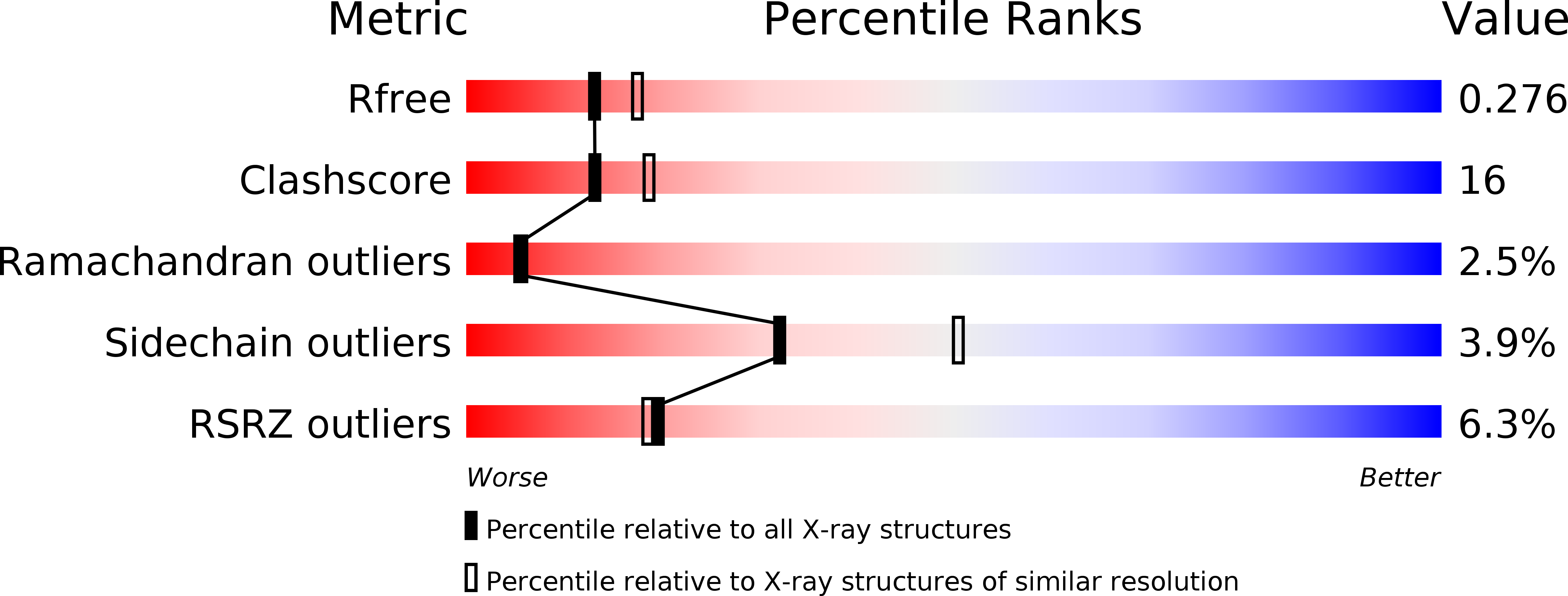
R-Value Free:
0.28
R-Value Work:
0.23
R-Value Observed:
0.23
Space Group:
C 2 2 21