1YUK image
Deposition Date 2005-02-14
Release Date 2005-07-19
Last Version Date 2024-11-13
Entry Detail
PDB ID:
1YUK
Keywords:
Title:
The crystal structure of the PSI/Hybrid domain/ I-EGF1 segment from the human integrin beta2 at 1.8 resolution
Biological Source:
Source Organism:
Homo sapiens (Taxon ID: 9606)
Host Organism:
Method Details:
Experimental Method:
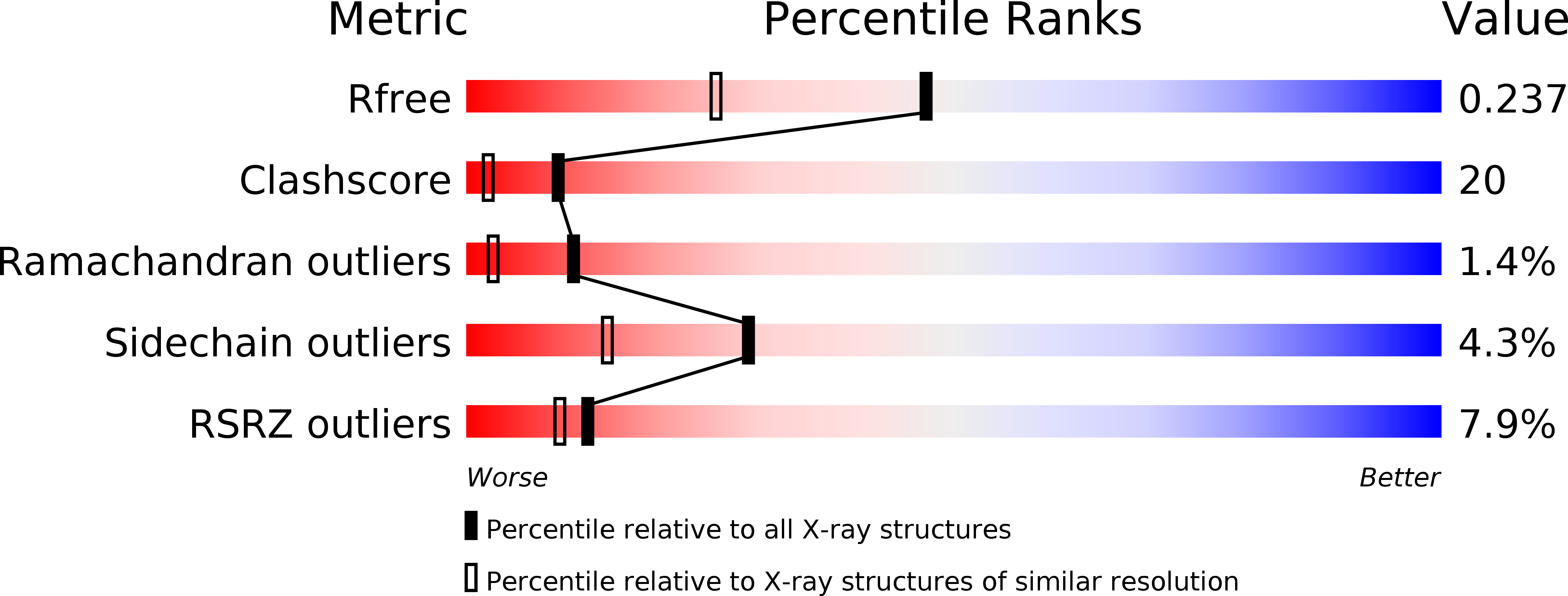
Resolution:
1.80 Å
R-Value Free:
0.25
R-Value Work:
0.23
R-Value Observed:
0.23
Space Group:
P 1 21 1
Macromolecular Entities
Structures with similar UniProt ID
Protein Blast
Polymer Type:polypeptide(L)
Molecule:Integrin beta-2 A chain
Gene (Uniprot):ITGB2
Chain IDs:A
Chain Length:103
Number of Molecules:1
Biological Source:Homo sapiens
Structures with similar UniProt ID
Protein Blast
Polymer Type:polypeptide(L)
Molecule:Integrin beta-2 B chain
Gene (Uniprot):ITGB2
Chain IDs:B
Chain Length:120
Number of Molecules:1
Biological Source:Homo sapiens
Ligand Molecules
Primary Citation
The Crystal Structure of the Plexin-Semaphorin-Integrin Domain/Hybrid Domain/I-EGF1 Segment from the Human Integrin {beta}2 Subunit at 1.8-A Resolution
J.Biol.Chem. 280 30586 30593 (2005)
PMID: 15965234 DOI: 10.1074/jbc.M502525200

Abstact

Integrins are modular (alphabeta) heterodimeric proteins that mediate cell adhesion and convey signals across the plasma membrane. Interdomain motions play a key role in signal transduction by propagating structural changes through the molecule, thus controlling the activation state and adhesive properties of the integrin. We expressed a soluble fragment of the human integrin beta2 subunit comprising the plexin-semaphorin-integrin domain (PSI)/hybrid domain/I-EGF1 fragment and present its crystal structure at 1.8-A resolution. The structure reveals an elongated molecule with a rigid architecture stabilized by nine disulfide bridges. The PSI domain is located centrally and participates in the formation of extended interfaces with the hybrid domain and I-EGF1 domains, respectively. The hybrid domain/PSI interface involves the burial of an Arg residue, and contacts between PSI and I-EGF1 are mainly mediated by well conserved Arg and Trp residues. Conservation of key interacting residues across the various integrin beta subunits sequences suggests that our structure represents a good model for the entire integrin family. Superposition with the integrin beta3 receptor in its bent conformation suggests that an articulation point is present at the linkage between its I-EGF1 and I-EGF2 modules and underlines the importance of this region for the control of integrin-mediated cell adhesion.

Legend

Protein

Chemical

Disease

Primary Citation of related structures