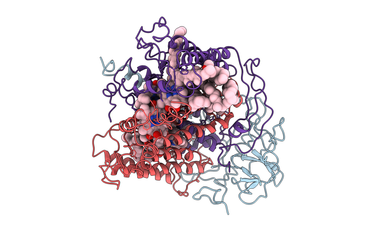
1YST image
Entry Detail
PDB ID:
1YST
Title:
STRUCTURE OF THE PHOTOCHEMICAL REACTION CENTER OF A SPHEROIDENE CONTAINING PURPLE BACTERIUM, RHODOBACTER SPHAEROIDES Y, AT 3 ANGSTROMS RESOLUTION
Biological Source:
Source Organism:
PDB Version:
Deposition Date:
1994-12-07
Release Date:
1995-02-27
Method Details:
Experimental Method:
Resolution:
3.00 Å
R-Value Work:
0.23
R-Value Observed:
0.23
Space Group:
P 21 21 21
Macromolecular Entities
Polymer Type:polypeptide(L)
Description:PHOTOSYNTHETIC REACTION CENTER (H SUBUNIT)
Chain IDs:C (auth: H)
Chain Length:260
Number of Molecules:1
Biological Source:Rhodobacter sphaeroides
Polymer Type:polypeptide(L)
Description:PHOTOSYNTHETIC REACTION CENTER (L SUBUNIT)
Chain IDs:A (auth: L)
Chain Length:273
Number of Molecules:1
Biological Source:Rhodobacter sphaeroides
Polymer Type:polypeptide(L)
Description:PHOTOSYNTHETIC REACTION CENTER (M SUBUNIT)
Chain IDs:B (auth: M)
Chain Length:305
Number of Molecules:1
Biological Source:Rhodobacter sphaeroides
Primary Citation
Structure of the photochemical reaction centre of a spheroidene-containing purple-bacterium, Rhodobacter sphaeroides Y, at 3 A resolution.
Acta Crystallogr.,Sect.D 51 368 379 (1995)
PMID: 15299304 DOI: 10.1107/S0907444994013120

Abstact

The crystal structure of the photochemical reaction centre from Rhodobacter sphaeroides Y, a carotenoid-containing wild-type purple bacterium, has been determined at 3 A resolution. This membrane complex consists of three subunits (281, 307 and 260 residues, respectively) and ten cofactors. It was crystallized in presence of beta-D-octylglucoside. The crystals are orthorhombic with unit-cell dimensions, a = 143.7, b = 139.8, c = 78.7 A, space group P2(1)2(1)2(1) with four molecules in the unit cell. Refinement of the structure by X-PLOR and manual reconstructions yielded an R value of 22.1% for 19630 reflections between 7 and 3 A. The secondary structure is highly homologous to those determined for Rhodopseudomonas viridis (Protein Data Bank entry 1PRC) and Rhodobacter sphaeroides R26 (Protein Data Bank entry 4RCR) reaction centres. In the latter two structures one Fe(2+) ion located between the two quinones is coordinated by four histidines and one glutamic acid. In the Rhodobacter sphaeroides Y structure, Mn(2+) occupies the same position with identical ligands and geometry. The carotenoid conformation which is a non-planar 15-15'-cis spheroidene molecule in our structure differs from the 13-14-cis 2,4-dihydroneusporene in the Rhodopseudomonas viridis structure.

Legend

Protein

Chemical

Disease

Primary Citation of related structures