1YSE image
Deposition Date 2005-02-08
Release Date 2006-01-03
Last Version Date 2024-05-29
Entry Detail
PDB ID:
1YSE
Title:
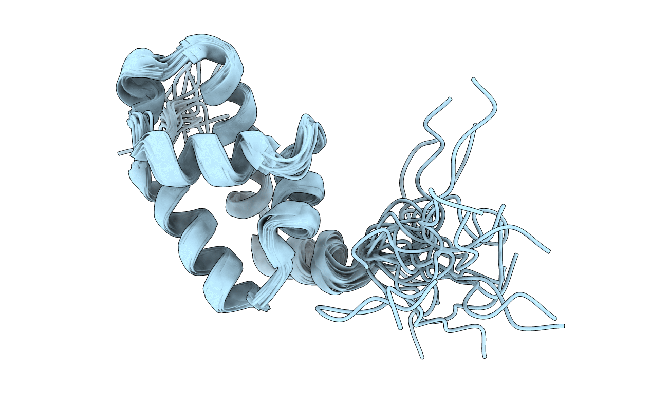
Solution structure of the MAR-binding domain of SATB1
Biological Source:
Source Organism:
Homo sapiens (Taxon ID: 9606)
Method Details:
Experimental Method:
Conformers Calculated:
50
Conformers Submitted:
20
Selection Criteria:
structures with the lowest energy
Macromolecular Entities
Polymer Type:polypeptide(L)
Molecule:DNA-binding protein SATB1
Gene (Uniprot):SATB1
Chain IDs:A
Chain Length:141
Number of Molecules:1
Biological Source:Homo sapiens
Ligand Molecules
Primary Citation
Solution Structure and DNA-binding Mode of the Matrix Attachment Region-binding Domain of the Transcription Factor SATB1 That Regulates the T-cell Maturation
J.Biol.Chem. 281 5319 5327 (2006)
PMID: 16371359 DOI: 10.1074/jbc.M510933200

Abstact

SATB1 is a transcriptional regulator controlling the gene expression that is essential in the maturation of the immune T-cell. SATB1 binds to the nuclear matrix attachment regions of DNA, where it recruits histone deacetylase and represses transcription through a local chromatin remodeling. Here we determined the solution structure of the matrix attachment region-binding domain, possessing similarity to the CUT DNA-binding domain, of human SATB1 by NMR spectroscopy. The structure consists of five alpha-helices, in which the N-terminal four are arranged similarly to the four-helix structure of the CUT domain of hepatocyte nuclear factor 6alpha. By an NMR chemical shift perturbation analysis and by surface plasmon resonance analyses of SATB1 mutant proteins, an interface for DNA binding was revealed to be located at the third helix and the surrounding regions. Surface plasmon resonance experiments using groove-specific binding drugs and methylated DNAs indicated that the domain recognizes DNA from the major groove side. These observations suggested that SATB1 possesses a DNA-binding mode similar to that of the POU-specific DNA-binding domain, which is known to share structural similarity to the four-helix CUT domain.

Legend

Protein

Chemical

Disease

Primary Citation of related structures