Deposition Date
2005-02-04
Release Date
2006-03-21
Last Version Date
2024-04-03
Entry Detail
PDB ID:
1YRR
Keywords:
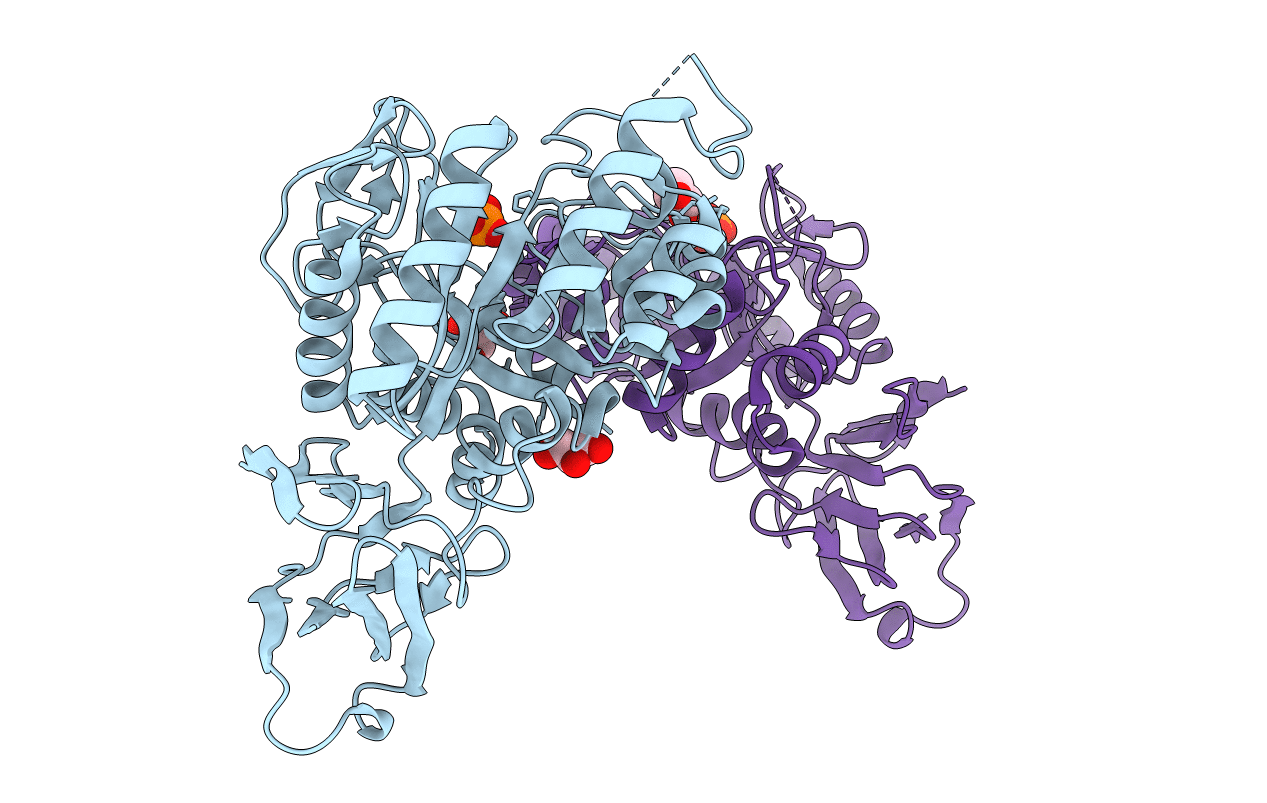
Title:
Crystal Structure Of The N-Acetylglucosamine-6-Phosphate Deacetylase From Escherichia Coli K12 at 2.0 A Resolution
Biological Source:
Source Organism(s):
Escherichia coli (Taxon ID: 562)
Expression System(s):
Method Details:
Experimental Method:
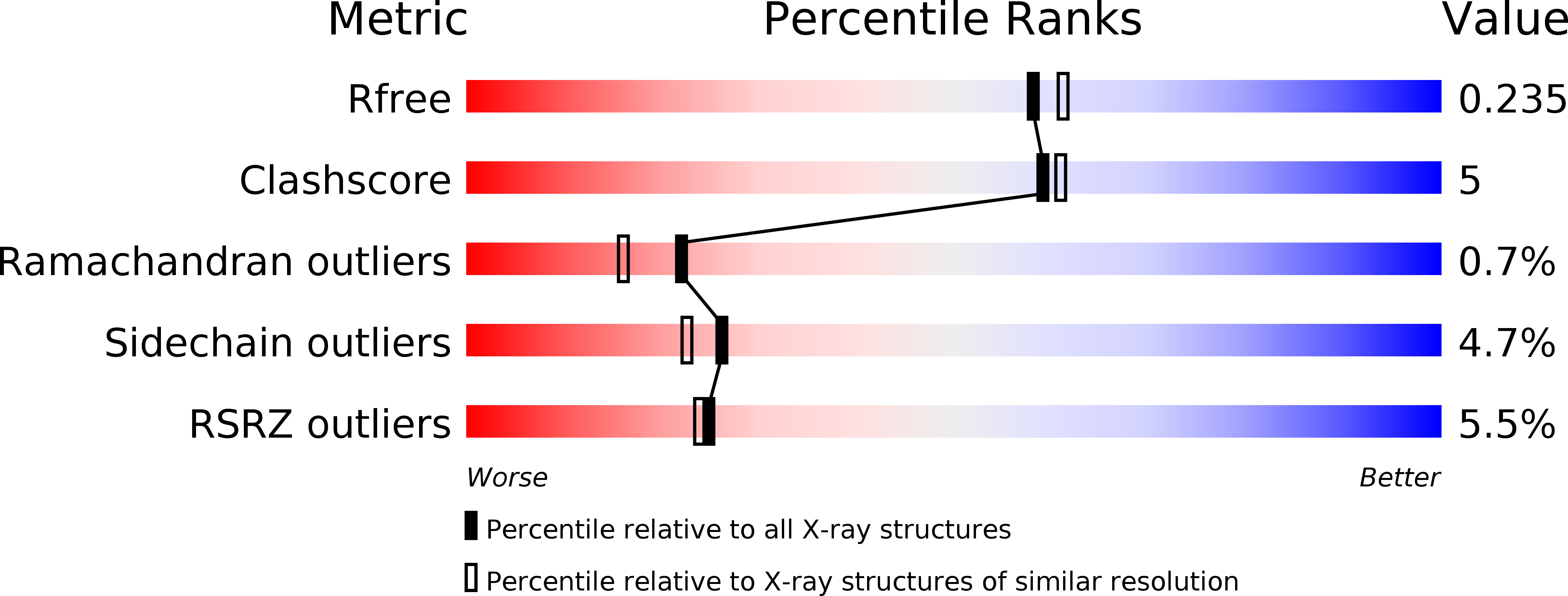
Resolution:
2.00 Å
R-Value Free:
0.19
R-Value Work:
0.15
R-Value Observed:
0.16
Space Group:
P 21 21 2