1YQ8 image
Deposition Date 2005-02-01
Release Date 2005-04-26
Last Version Date 2024-03-13
Entry Detail
PDB ID:
1YQ8
Keywords:
Title:
PRD1 vertex protein P5
Biological Source:
Source Organism:
Host Organism:
Method Details:
Experimental Method:
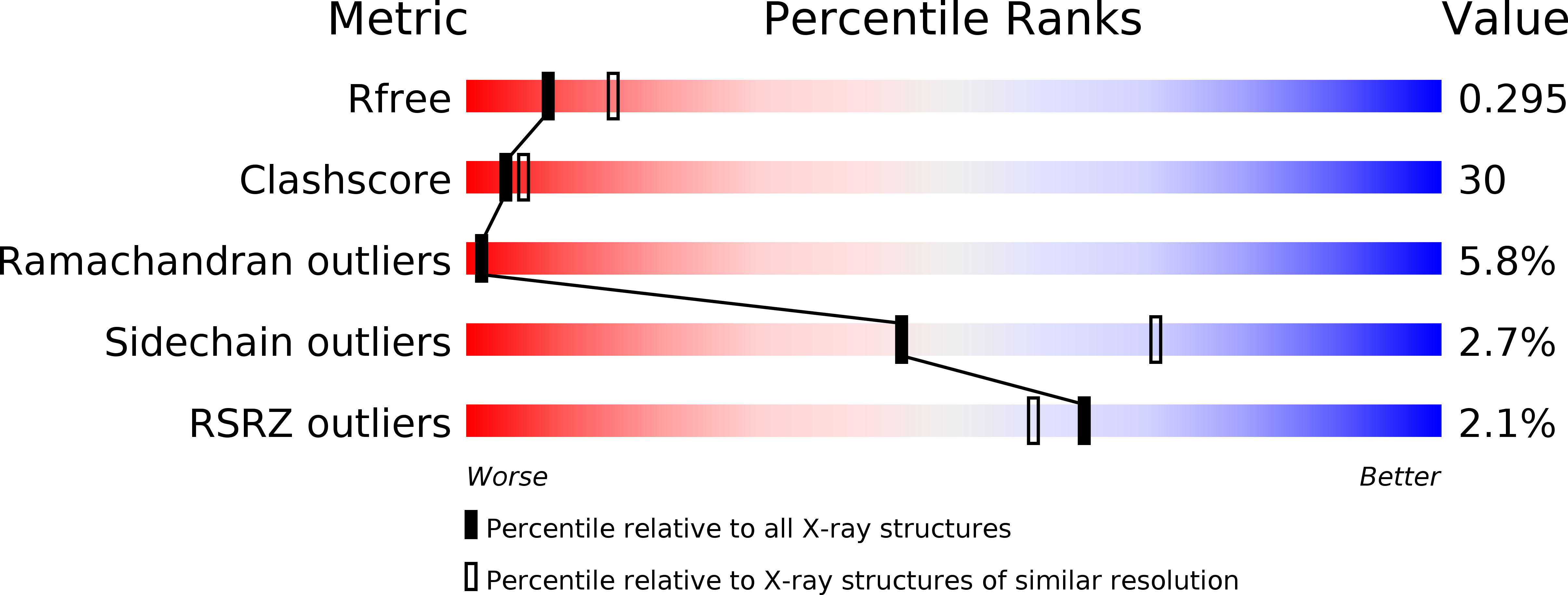
Resolution:
2.60 Å
R-Value Free:
0.3
R-Value Work:
0.26
R-Value Observed:
0.26
Space Group:
H 3
Macromolecular Entities
Polymer Type:polypeptide(L)
Molecule:Minor capsid protein
Gene (Uniprot):V
Mutations:deletion mutant
Chain IDs:A
Chain Length:191
Number of Molecules:1
Biological Source:Enterobacteria phage PRD1
Ligand Molecules
Primary Citation
The structure of the bacteriophage PRD1 spike sheds light on the evolution of viral capsid architecture.
Mol.Cell 18 161 170 (2005)
PMID: 15837420 DOI: 10.1016/j.molcel.2005.03.019

Abstact

Comparisons of bacteriophage PRD1 and adenovirus protein structures and virion architectures have been instrumental in unraveling an evolutionary relationship and have led to a proposal of a phylogeny-based virus classification. The structure of the PRD1 spike protein P5 provides further insight into the evolution of viral proteins. The crystallized P5 fragment comprises two structural domains: a globular knob and a fibrous shaft. The head folds into a ten-stranded jelly roll beta barrel, which is structurally related to the tumor necrosis factor (TNF) and the PRD1 coat protein domains. The shaft domain is a structural counterpart to the adenovirus spike shaft. The structural relationships between PRD1, TNF, and adenovirus proteins suggest that the vertex proteins may have originated from an ancestral TNF-like jelly roll coat protein via a combination of gene duplication and deletion.

Legend

Protein

Chemical

Disease

Primary Citation of related structures