1YO8 image
Deposition Date 2005-01-26
Release Date 2005-09-27
Last Version Date 2024-11-20
Entry Detail
PDB ID:
1YO8
Keywords:
Title:
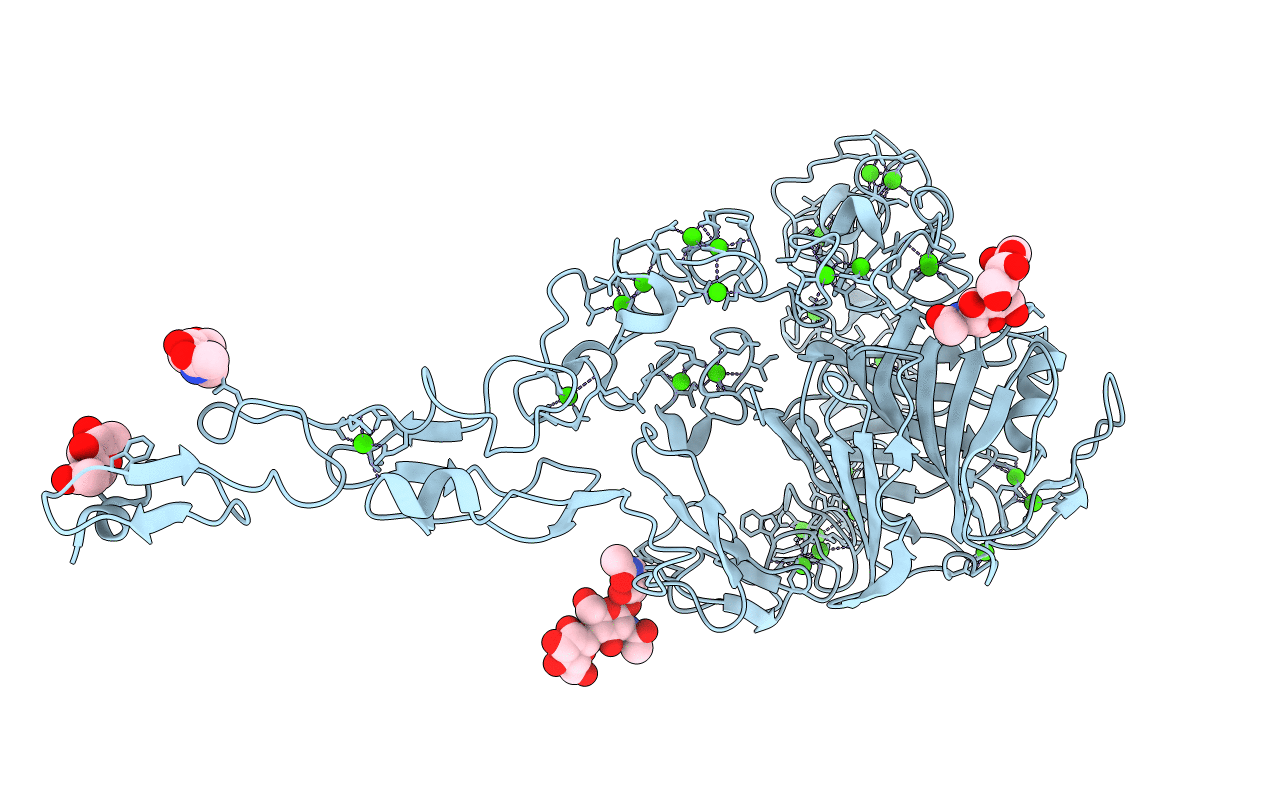
Structure of the C-terminal domain of human thrombospondin-2
Biological Source:
Source Organism:
Homo sapiens (Taxon ID: 9606)
Host Organism:
Method Details:
Experimental Method:
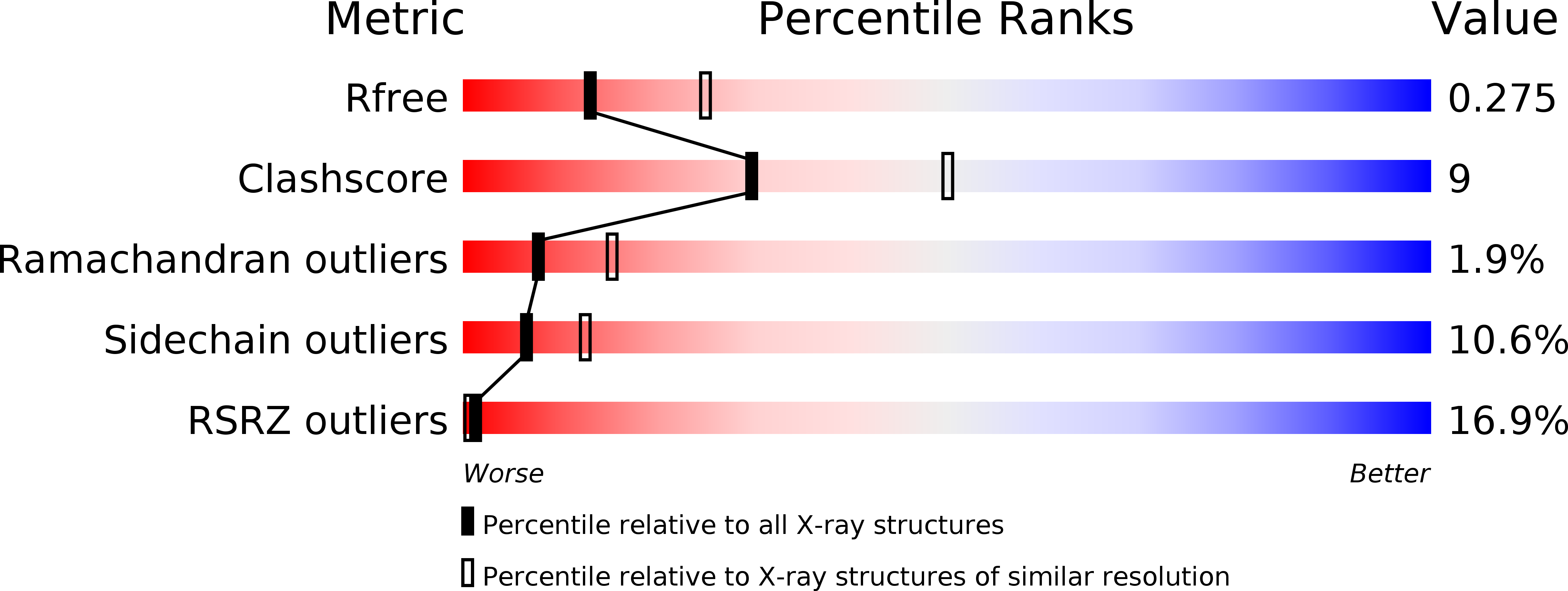
Resolution:
2.60 Å
R-Value Free:
0.28
R-Value Work:
0.21
R-Value Observed:
0.22
Space Group:
I 2 2 2
Macromolecular Entities
Structures with similar UniProt ID
Protein Blast
Polymer Type:polypeptide(L)
Molecule:thrombospondin-2
Gene (Uniprot):THBS2
Chain IDs:A
Chain Length:634
Number of Molecules:1
Biological Source:Homo sapiens
Modified Residue
Compound ID Chain ID Parent Comp ID Details 2D Image
ASN A ASN GLYCOSYLATION SITE
MSE A MET SELENOMETHIONINE
SER A SER GLYCOSYLATION SITE
Primary Citation
Structure of the calcium-rich signature domain of human thrombospondin-2
Nat.Struct.Mol.Biol. 12 910 914 (2005)
PMID: 16186819 DOI: 10.1038/nsmb997

Abstact

Thrombospondins (THBSs) are secreted glycoproteins that have key roles in interactions between cells and the extracellular matrix. Here, we describe the 2.6-A-resolution crystal structure of the glycosylated signature domain of human THBS2, which includes three epidermal growth factor-like modules, 13 aspartate-rich repeats and a lectin-like module. These elements interact extensively to form three structural regions termed the stalk, wire and globe. The THBS2 signature domain is stabilized by these interactions and by a network of 30 bound Ca(2+) ions and 18 disulfide bonds. The structure suggests how genetic alterations of THBSs result in disease.

Legend

Protein

Chemical

Disease

Primary Citation of related structures
Feedback Form
Name
Email
Institute
Feedback