Deposition Date
2005-01-21
Release Date
2005-05-31
Last Version Date
2024-11-06
Entry Detail
PDB ID:
1YMH
Keywords:
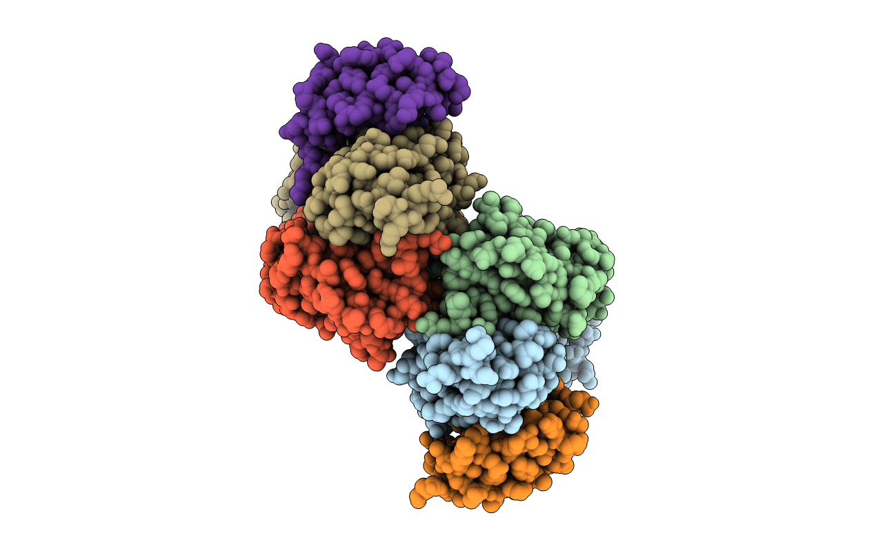
Title:
anti-HCV Fab 19D9D6 complexed with protein L (PpL) mutant A66W
Biological Source:
Source Organism(s):
Finegoldia magna (Taxon ID: 334413)
Mus musculus (Taxon ID: 10090)
Mus musculus (Taxon ID: 10090)
Expression System(s):
Method Details:
Experimental Method:
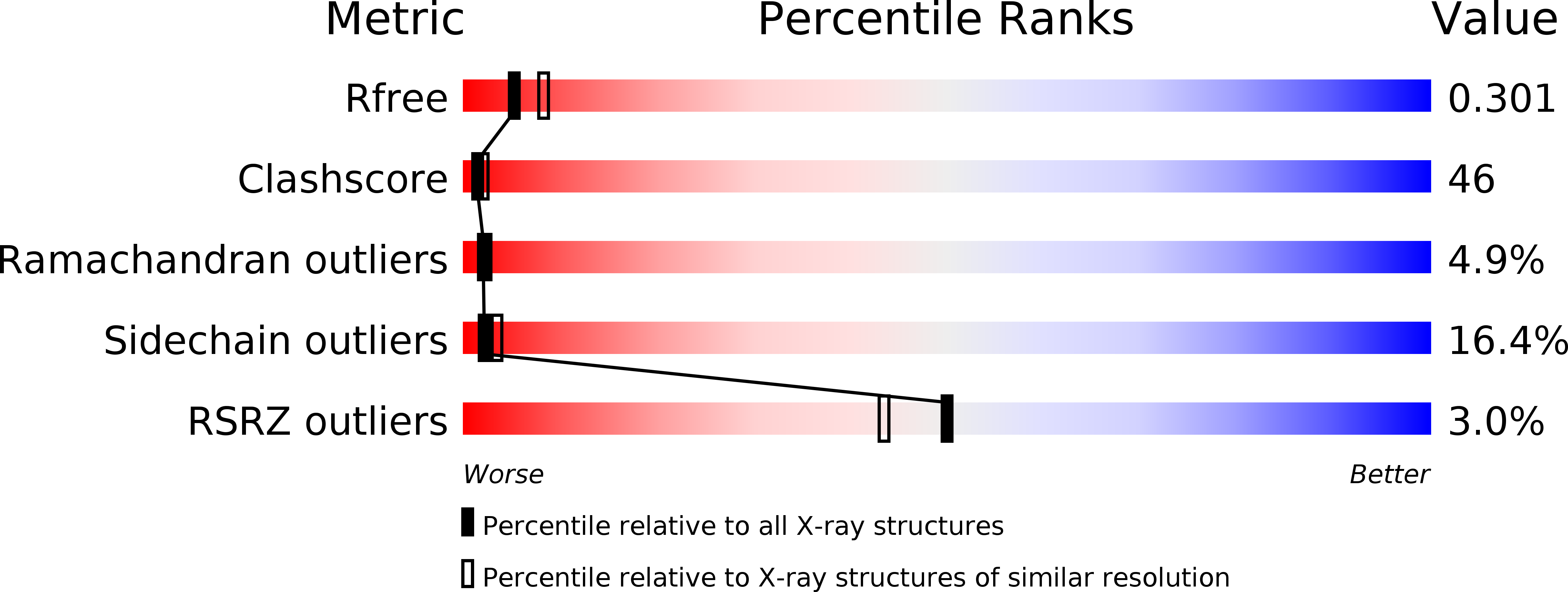
Resolution:
2.60 Å
R-Value Free:
0.30
R-Value Work:
0.22
R-Value Observed:
0.22
Space Group:
P 21 21 21