
Deposition Date
2005-01-19
Release Date
2005-06-28
Last Version Date
2023-11-15
Entry Detail
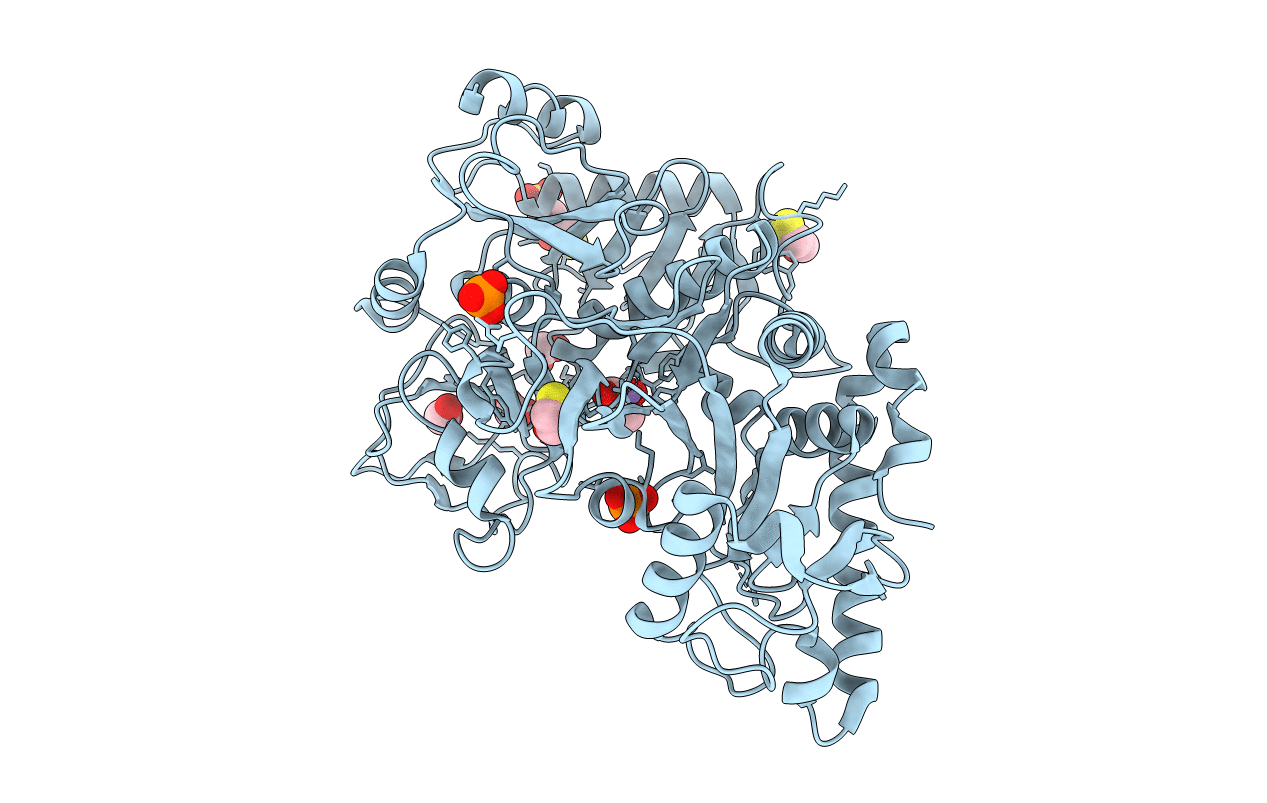
PDB ID:
1YLH
Keywords:
Title:
Crystal Structure of Phosphoenolpyruvate Carboxykinase from Actinobaccilus succinogenes in Complex with Manganese and Pyruvate
Biological Source:
Source Organism(s):
Actinobacillus succinogenes (Taxon ID: 67854)
Expression System(s):
Method Details:
Experimental Method:
Resolution:
1.70 Å
R-Value Free:
0.21
R-Value Work:
0.19
R-Value Observed:
0.19
Space Group:
P 1 21 1
