1YKN image
Deposition Date 2005-01-18
Release Date 2005-08-16
Last Version Date 2024-10-30
Entry Detail
PDB ID:
1YKN
Keywords:
Title:
Protocatechuate 3,4-dioxygenase Y408E mutant bound to DHB
Biological Source:
Source Organism:
Host Organism:
Method Details:
Experimental Method:
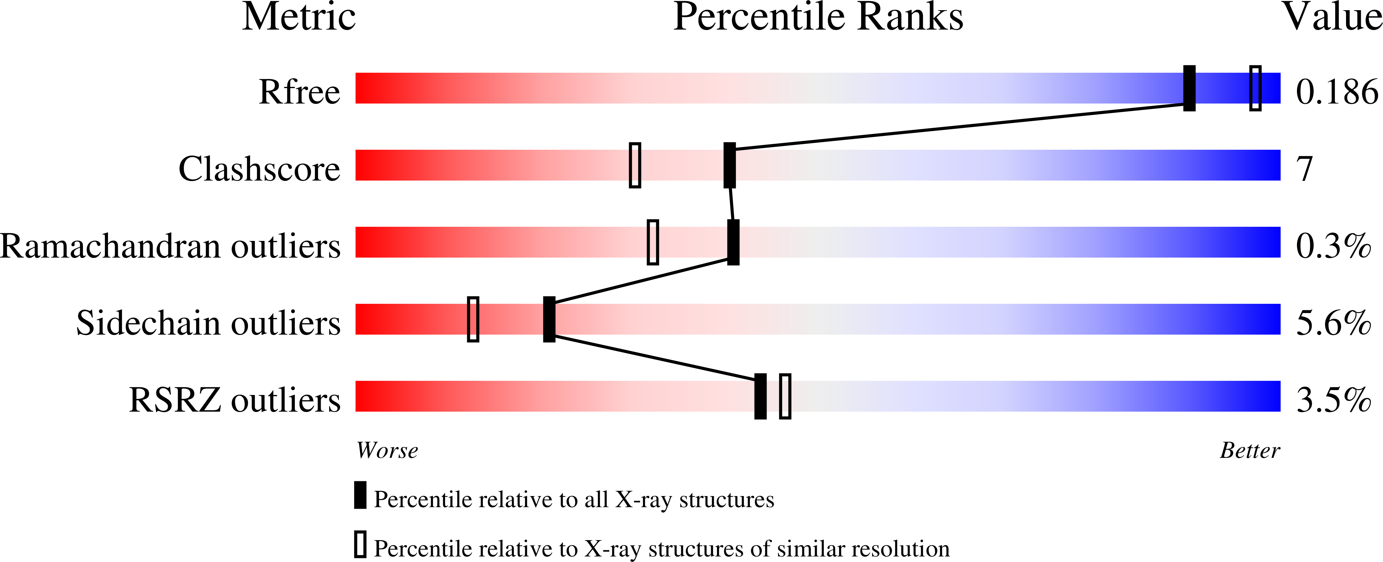
Resolution:
2.06 Å
R-Value Free:
0.19
R-Value Work:
0.14
R-Value Observed:
0.14
Space Group:
I 1 2 1
Macromolecular Entities
Polymer Type:polypeptide(L)
Molecule:Protocatechuate 3,4-dioxygenase alpha chain
Gene (Uniprot):pcaG
Chain IDs:A, C, E, G, I, K
Chain Length:200
Number of Molecules:6
Biological Source:Pseudomonas putida
Polymer Type:polypeptide(L)
Molecule:Protocatechuate 3,4-dioxygenase beta chain
Gene (Uniprot):pcaH
Mutations:Y408E
Chain IDs:B, D, F, H, J, L
Chain Length:238
Number of Molecules:6
Biological Source:Pseudomonas putida
Modified Residue
Compound ID Chain ID Parent Comp ID Details 2D Image
CME B CYS S,S-(2-HYDROXYETHYL)THIOCYSTEINE
Primary Citation
Roles of the equatorial tyrosyl iron ligand of protocatechuate 3,4-dioxygenase in catalysis
Biochemistry 44 11024 11039 (2005)
PMID: 16101286 DOI: 10.1021/bi050902i

Abstact

The active site Fe(III) of protocatechuate 3,4-dioxygenase (3,4-PCD) from Pseudomonas putida is ligated axially by Tyr447 and His462 and equatorially by Tyr408, His460, and OH(-). Tyr447 and OH(-) are displaced as protocatechuate (3,4-dihydroxybenzoate, PCA) chelates the iron and appear to serve as in situ bases to promote this process. The role(s) of Tyr408 is (are) explored here using mutant enzymes that exhibit less than 0.1% wild-type activity. The X-ray crystal structures of the mutants and their PCA complexes show that the new shorter residues in the 408 position cannot ligate the iron and instead interact with the iron through solvents. Moreover, PCA binds as a monodentate rather than a bidentate ligand, and Tyr447 fails to dissociate. Although the new residues at position 408 do not directly bind to the iron, large changes in the spectroscopic and catalytic properties are noted among the mutant enzymes. Resonance Raman features show that the Fe-O bond of the monodentate 4-hydroxybenzoate (4HB) inhibitor complex is significantly stronger in the mutants than in wild-type 3,4-PCD. Transient kinetic studies show that PCA and 4HB bind to 3,4-PCD in a fast, reversible step followed by a step in which coordination to the metal occurs; the latter process is at least 50-fold slower in the mutant enzymes. It is proposed that, in wild-type 3,4-PCD, the Lewis base strength of Tyr408 lowers the Lewis acidity of the iron to foster the rapid exchange of anionic ligands during the catalytic cycle. Accordingly, the increase in Lewis acidity of the iron caused by substitution of this residue by solvent tends to make the iron substitution inert. Tyr447 cannot be released to allow formation of the usual dianionic PCA chelate complex with the active site iron, and the rate of electrophilic attack by O(2) becomes rate limiting overall. The structures of the PCA complexes of these mutant enzymes show that hydrogen-bonding interactions between the new solvent ligand and the new second-sphere residue in position 408 allow this residue to significantly influence the spectroscopic and kinetic properties of the enzymes.

Legend

Protein

Chemical

Disease

Primary Citation of related structures