1YKD image
Deposition Date 2005-01-17
Release Date 2005-02-22
Last Version Date 2024-02-14
Entry Detail
PDB ID:
1YKD
Keywords:
Title:
Crystal Structure of the Tandem GAF Domains from a Cyanobacterial Adenylyl Cyclase: Novel Modes of Ligand-Binding and Dimerization
Biological Source:
Source Organism:
Anabaena sp. (Taxon ID: 1167)
Host Organism:
Method Details:
Experimental Method:
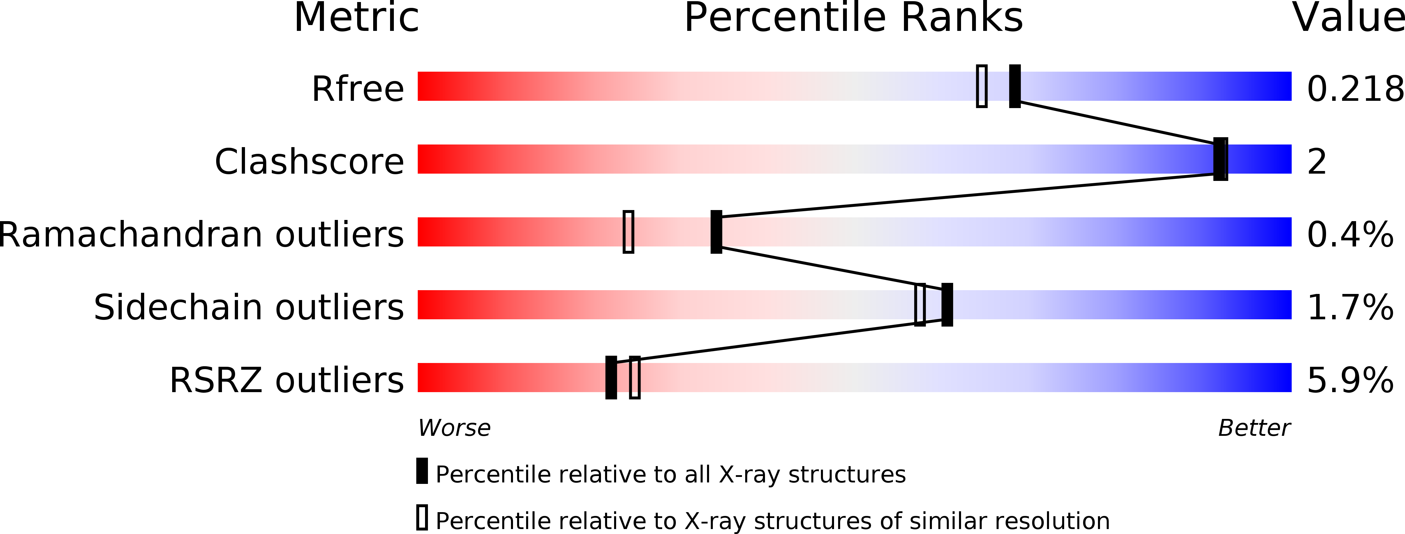
Resolution:
1.90 Å
R-Value Free:
0.21
R-Value Work:
0.18
R-Value Observed:
0.18
Space Group:
P 1
Macromolecular Entities
Polymer Type:polypeptide(L)
Molecule:adenylate cyclase
Gene (Uniprot):cyaB2
Chain IDs:A, B
Chain Length:398
Number of Molecules:2
Biological Source:Anabaena sp.
Ligand Molecules
Primary Citation
Crystal structure of the tandem GAF domains from a cyanobacterial adenylyl cyclase: Modes of ligand binding and dimerization
Proc.Natl.Acad.Sci.USA 102 3082 3087 (2005)
PMID: 15708973 DOI: 10.1073/pnas.0409913102

Abstact

In several species, GAF domains, which are widely expressed small-molecule-binding domains that regulate enzyme activity, are known to bind cyclic nucleotides. However, the molecular mechanism by which cyclic nucleotide binding affects enzyme activity is not known for any GAF domain. In the cyanobacterium, Anabaena, the cyaB1 and cyaB2 genes encode adenylyl cyclases that are stimulated by binding of cAMP to their N-terminal GAF domains. Replacement of the tandem GAF-A/B domains in cyaB1 with the mammalian phosphodiesterase 2A GAF-A/B tandem domains allows regulation of the chimeric protein by cGMP, suggesting a highly conserved mechanism of activation. Here, we describe the 1.9-A crystal structure of the tandem GAF-A/B domains of cyaB2 with bound cAMP and compare it to the previously reported structure of the PDE2A GAF-A/B. Unexpectedly, the cyaB2 GAF-A/B dimer is antiparallel, unlike the parallel dimer of PDE2A. Moreover, there is clear electron density for cAMP in both GAF-A and -B, whereas in PDE2A, cGMP is found only in GAF-B. Phosphate and ribose group contacts are similar to those in PDE2A. However, the purine-binding pockets appear very different from that in PDE2A GAF-B. Differences in the beta2-beta3 loop suggest that this loop confers much of the ligand specificity in this and perhaps in many other GAF domains. Finally, a conserved asparagine appears to be a new addition to the signature NKFDE motif, and a mechanism for this motif to stabilize the cNMP-binding pocket is proposed.

Legend

Protein

Chemical

Disease

Primary Citation of related structures