1YJG image
Entry Detail
PDB ID:
1YJG
Keywords:
Title:
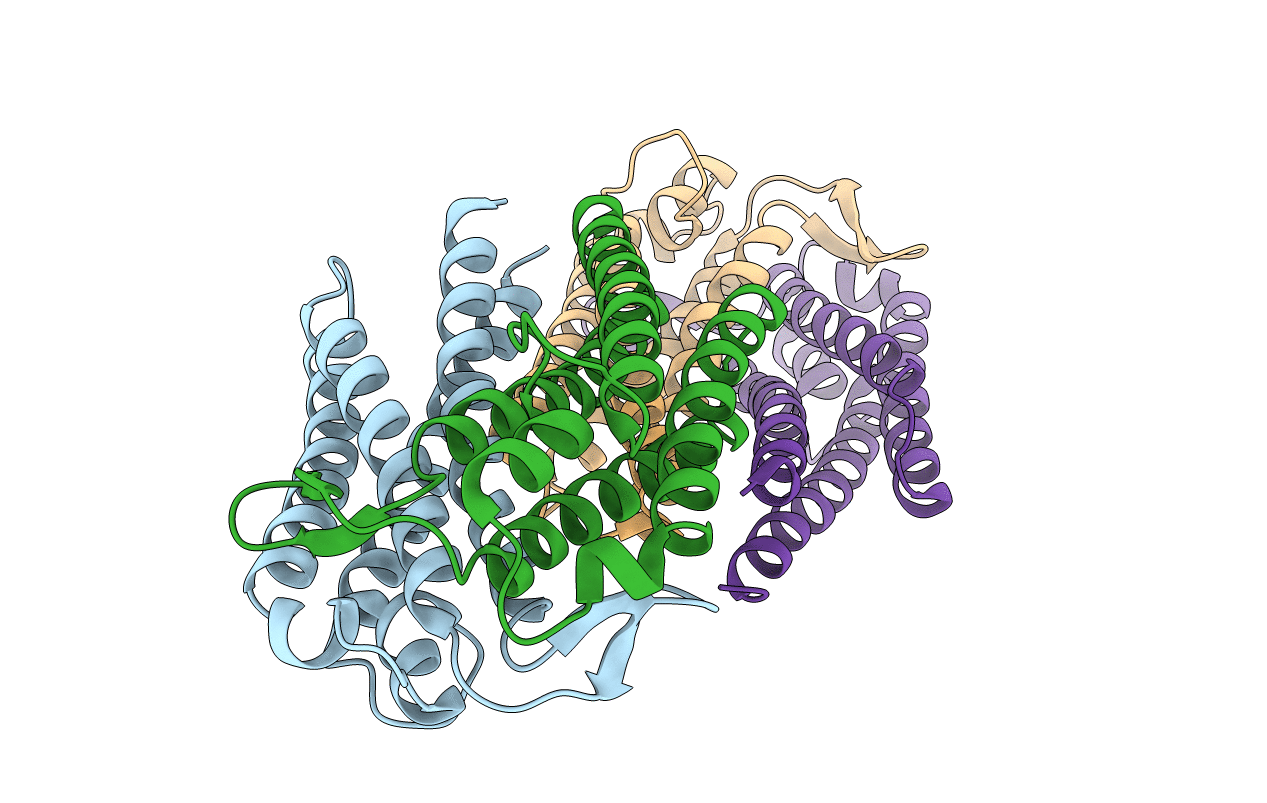
Variable Small Protein 1 of Borrelia turicatae (VspA or Vsp1)
Biological Source:
Source Organism:
PDB Version:
Deposition Date:
2005-01-14
Release Date:
2006-01-24
Method Details:
Experimental Method:
Resolution:
2.22 Å
R-Value Free:
0.26
R-Value Work:
0.20
R-Value Observed:
0.20
Space Group:
C 1 2 1
Macromolecular Entities
Polymer Type:polypeptide(L)
Description:Surface protein VspA
Chain IDs:A, B, C (auth: D), D (auth: E)
Chain Length:158
Number of Molecules:4
Biological Source:Borrelia turicatae
Primary Citation
Crystal structure of neurotropism-associated variable surface protein 1 (Vsp1) of Borrelia turicatae.
J.Bacteriol. 188 4522 4530 (2006)
PMID: 16740958 DOI: 10.1128/JB.00028-06

Abstact

Vsp surface lipoproteins are serotype-defining antigens of relapsing fever spirochetes that undergo multiphasic antigenic variation to allow bacterial persistence in spite of an immune response. Two isogenic serotypes of Borrelia turicatae strain Oz1 differ in their Vsp sequences and in disease manifestations in infected mice: Vsp1 is associated with the selection of a neurological niche, while Vsp2 is associated with blood and skin infection. We report here crystal structures of the Vsp1 dimer at 2.7 and 2.2 A. The structures confirm that relapsing fever Vsp proteins share a common helical fold with OspCs of Lyme disease-causing Borrelia. The fold features an inner stem formed by highly conserved N and C termini and an outer "dome" formed by the variable central residues. Both Vsp1 and OspC structures possess small water-filled cavities, or pockets, that are lined largely by variable residues and are thus highly variable in shape. These features appear to signify tolerance of the Vsp-OspC fold for imperfect packing of residues at its antigenic surface. Structural comparison of Vsp1 with a homology model for Vsp2 suggests that observed differences in disease manifestation may arise in part from distinct differences in electrostatic surface properties; additional predicted positively charged surface patches on Vsp2 compared to Vsp1 may be sufficient to explain the relative propensity of Vsp2 to bind to acidic glycosaminoglycans.

Legend

Protein

Chemical

Disease

Primary Citation of related structures