1YIV image
Deposition Date 2005-01-13
Release Date 2005-07-26
Last Version Date 2023-10-25
Entry Detail
PDB ID:
1YIV
Keywords:
Title:
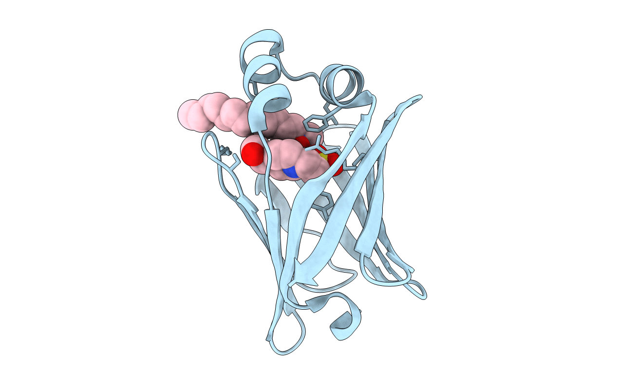
Structure of myelin P2 protein from Equine spinal cord
Biological Source:
Source Organism:
Equus caballus (Taxon ID: 9796)
Method Details:
Experimental Method:
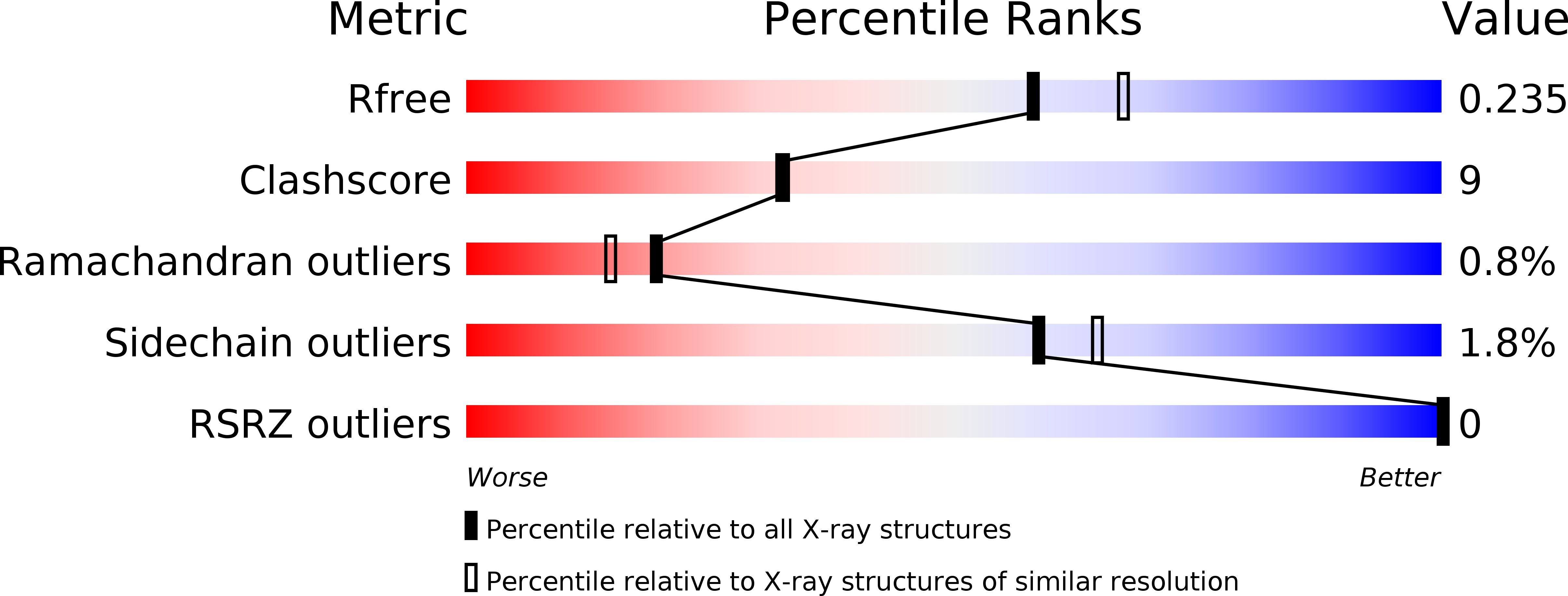
Resolution:
2.10 Å
R-Value Free:
0.23
R-Value Work:
0.18
R-Value Observed:
0.18
Space Group:
P 32 2 1
Macromolecular Entities
Polymer Type:polypeptide(L)
Molecule:Myelin P2 protein
Gene (Uniprot):PMP2
Chain IDs:A
Chain Length:131
Number of Molecules:1
Biological Source:Equus caballus
Primary Citation
Structure of myelin P2 protein from equine spinal cord.
Acta Crystallogr.,Sect.D 61 1067 1071 (2005)
PMID: 16041071 DOI: 10.1107/S0907444905014162

Abstact

Equine P2 protein has been isolated from horse spinal cord and its structure determined to 2.1 A. Since equine myelin is a viable alternative to bovine tissue for large-scale preparations, characterization of the proteins from equine spinal cord myelin has been initiated. There is an unusually high amount of P2 protein in equine CNS myelin compared with other species. The structure was determined by molecular replacement and subsequently refined to an R value of 0.187 (Rfree=0.233). The structure contains a molecule of the detergent LDAO and HEPES buffer in the binding cavity and is otherwise analogous to other cellular retinol-binding proteins.

Legend

Protein

Chemical

Disease

Primary Citation of related structures