1YIQ image
Deposition Date 2005-01-12
Release Date 2005-08-16
Last Version Date 2024-11-20
Entry Detail
PDB ID:
1YIQ
Keywords:
Title:
Molecular cloning and structural analysis of quinohemoprotein alcohol dehydrogenase ADHIIG from Pseudomonas putida HK5. Compariison to the other quinohemoprotein alcohol dehydrogenase ADHIIB found in the same microorganism.
Biological Source:
Source Organism:
Method Details:
Experimental Method:
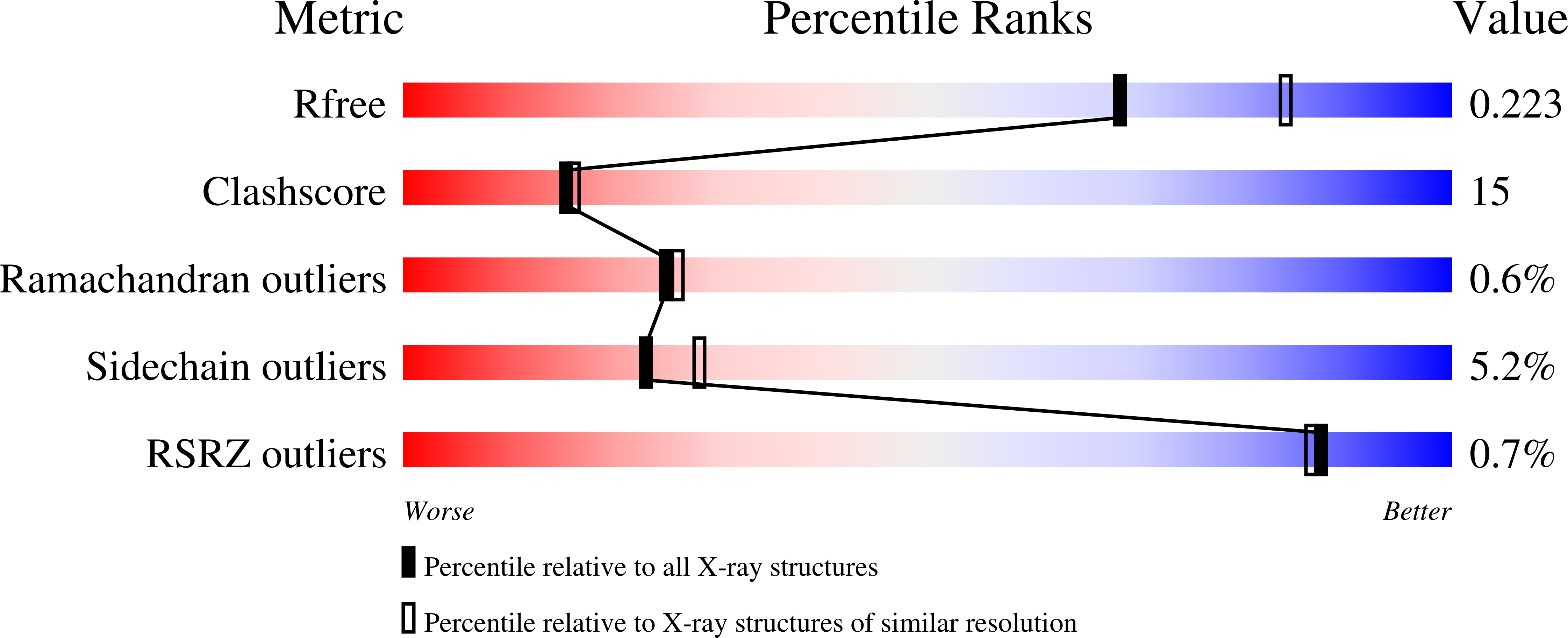
Resolution:
2.20 Å
R-Value Free:
0.22
R-Value Work:
0.17
Space Group:
P 61
Macromolecular Entities
Polymer Type:polypeptide(L)
Molecule:Quinohemoprotein alcohol dehydrogenase
Gene (Uniprot):qgdA
Chain IDs:A
Chain Length:689
Number of Molecules:1
Biological Source:Pseudomonas putida
Primary Citation
Molecular cloning and structural analysis of quinohemoprotein alcohol dehydrogenase ADH-IIG from Pseudomonas putida HK5
J.Mol.Biol. 352 91 104 (2005)
PMID: 16061256 DOI: 10.1016/j.jmb.2005.06.078

Abstact

Depending on the alcohols used as growth substrates, Pseudomonas putida HK5 produces two distinct quinohemoprotein alcohol dehydrogenases, ADH-IIB and ADH-IIG, both of which contain pyrroloquinoline quinone (PQQ) and heme c as the prosthetic groups but show different substrate specificities, especially for diol substrates. Molecular cloning of the gene of ADH-IIB and its crystal structure are already reported. Here, molecular cloning of the gene, qgdA, and solution of the three-dimensional structure of ADH-IIG are reported. The enzyme consists of 718 amino acid residues including a signal sequence of 29 amino acid residues. The PQQ domain is highly homologous to other quinoproteins, especially to quinohemoproteins. The crystal structure of ADH-IIG, determined at 2.2A resolution, shows that the overall structure and the amino acid residues involved in PQQ binding are quite similar to ADH-IIB and to another quinohemoprotein ADH, qhEDH from Comamonas testosteroni. However, the lengths of the linker regions connecting the PQQ and the cytochrome domains are different from each other, leading to a significant difference in orientation of the cytochrome domain with respect to the PQQ domain. Apart from ADH-IIB and qhEDH, ADH-IIG has an extra 12-residue helix within loop 3 in the PQQ domain and an extra 3(10) helix in the C terminus of the cytochrome domain, and both helices appear parallel and linked by a hydrogen bond. The amino acid residues contacting substrate/product in the crystal structures are also different among them. In the crystal structure of ADH-IIG with 1,2-propanediol, one of the hydroxyl groups of the substrate forms a hydrogen bond with O5 of PQQ and OD1 of Asp300, and the other interacts with a water molecule and with NE2 of Trp386, the corresponding residue of which is not found in ADH-IIB and qhEDH, and might be the residue responsible for making ADH-IIG prefer diol substrates.

Legend

Protein

Chemical

Disease

Primary Citation of related structures