1YI5 image
Deposition Date 2005-01-11
Release Date 2005-05-17
Last Version Date 2024-10-30
Entry Detail
PDB ID:
1YI5
Keywords:
Title:
Crystal structure of the a-cobratoxin-AChBP complex
Biological Source:
Source Organism:
Method Details:
Experimental Method:
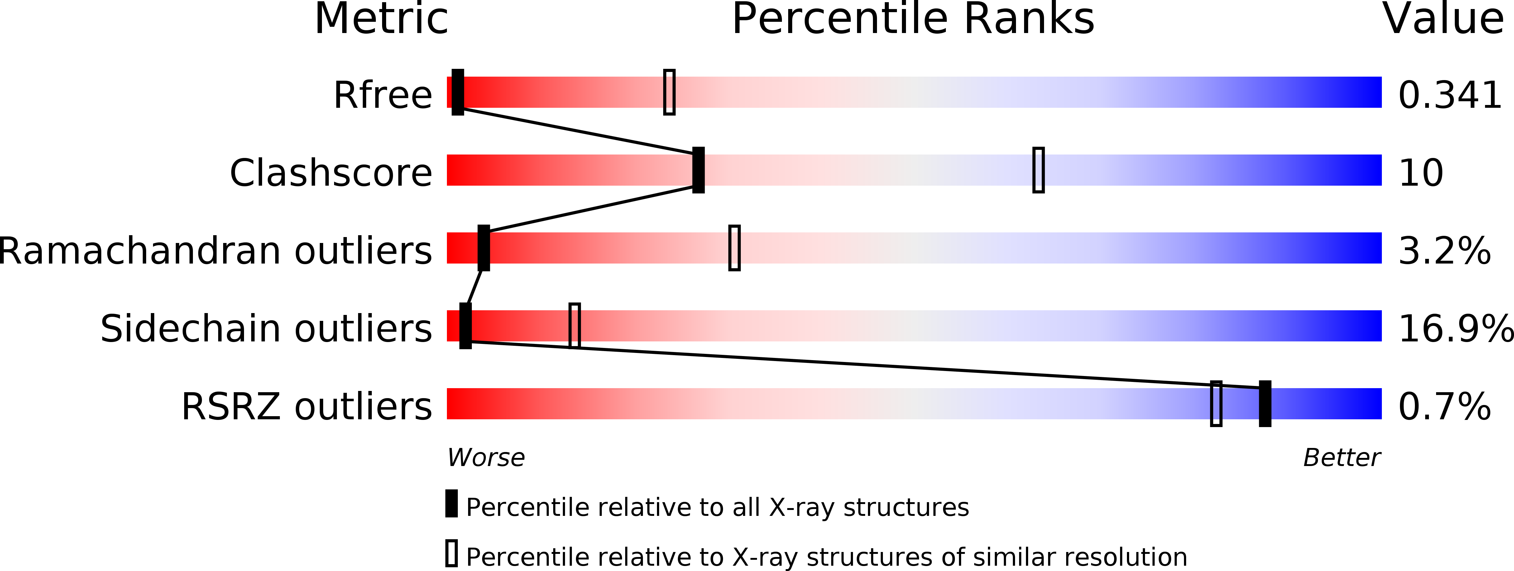
Resolution:
4.20 Å
R-Value Free:
0.37
R-Value Work:
0.33
R-Value Observed:
0.33
Space Group:
C 2 2 21
Macromolecular Entities
Structures with similar UniProt ID
Protein Blast
Polymer Type:polypeptide(L)
Molecule:Acetylcholine-binding protein
Chain IDs:A, C (auth: B), E (auth: C), G (auth: D), I (auth: E)
Chain Length:210
Number of Molecules:5
Biological Source:Lymnaea stagnalis
Structures with similar UniProt ID
Protein Blast
Polymer Type:polypeptide(L)
Molecule:Long neurotoxin 1
Chain IDs:B (auth: F), D (auth: G), F (auth: H), H (auth: I), J
Chain Length:71
Number of Molecules:5
Biological Source:Naja siamensis
Ligand Molecules
Primary Citation
Crystal structure of a Cbtx-AChBP complex reveals essential interactions between snake alpha-neurotoxins and nicotinic receptors
Embo J. 24 1512 1522 (2005)
PMID: 15791209 DOI: 10.1038/sj.emboj.7600620

Abstact

The crystal structure of the snake long alpha-neurotoxin, alpha-cobratoxin, bound to the pentameric acetylcholine-binding protein (AChBP) from Lymnaea stagnalis, was solved from good quality density maps despite a 4.2 A overall resolution. The structure unambiguously reveals the positions and orientations of all five three-fingered toxin molecules inserted at the AChBP subunit interfaces and the conformational changes associated with toxin binding. AChBP loops C and F that border the ligand-binding pocket move markedly from their original positions to wrap around the tips of the toxin first and second fingers and part of its C-terminus, while rearrangements also occur in the toxin fingers. At the interface of the complex, major interactions involve aromatic and aliphatic side chains within the AChBP binding pocket and, at the buried tip of the toxin second finger, conserved Phe and Arg residues that partially mimic a bound agonist molecule. Hence this structure, in revealing a distinctive and unpredicted conformation of the toxin-bound AChBP molecule, provides a lead template resembling a resting state conformation of the nicotinic receptor and for understanding selectivity of curaremimetic alpha-neurotoxins for the various receptor species.

Legend

Protein

Chemical

Disease

Primary Citation of related structures