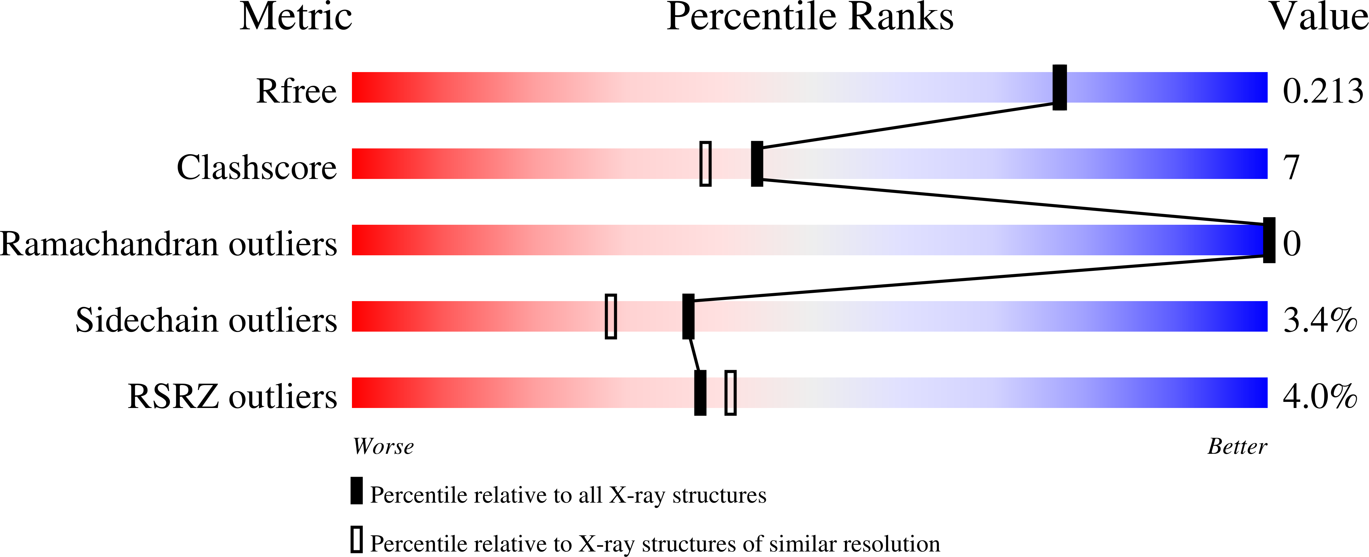
1YFD image
Deposition Date 2004-12-31
Release Date 2005-02-15
Last Version Date 2023-08-23
Entry Detail
PDB ID:
1YFD
Keywords:
Title:
Crystal structure of the Y122H mutant of ribonucleotide reductase R2 protein from E. coli
Biological Source:
Source Organism:
Escherichia coli (Taxon ID: 562)
Host Organism:
Method Details:
Experimental Method:
Resolution:
1.90 Å
R-Value Free:
0.20
R-Value Work:
0.17
R-Value Observed:
0.17
Space Group:
P 21 21 21
Macromolecular Entities
Polymer Type:polypeptide(L)
Molecule:Ribonucleoside-diphosphate reductase 1 beta chain
Gene (Uniprot):nrdB
Mutations:Y122H
Chain IDs:A, B
Chain Length:375
Number of Molecules:2
Biological Source:Escherichia coli
Primary Citation
A new tyrosyl radical on Phe208 as ligand to the diiron center in Escherichia coli ribonucleotide reductase, mutant R2-Y122H. Combined x-ray diffraction and EPR/ENDOR studies
J.Biol.Chem. 280 11233 11246 (2005)
PMID: 15634667 DOI: 10.1074/jbc.M414634200

Abstact

The R2 protein subunit of class I ribonucleotide reductase (RNR) belongs to a structurally related family of oxygen bridged diiron proteins. In wild-type R2 of Escherichia coli, reductive cleavage of molecular oxygen by the diferrous iron center generates a radical on a nearby tyrosine residue (Tyr122), which is essential for the enzymatic activity of RNR, converting ribonucleotides into deoxyribonucleotides. In this work, we characterize the mutant E. coli protein R2-Y122H, where the radical site is substituted with a histidine residue. The x-ray structure verifies the mutation. R2-Y122H contains a novel stable paramagnetic center which we name H, and which we have previously proposed to be a diferric iron center with a strongly coupled radical, Fe(III)Fe(III)R.. Here we report a detailed characterization of center H, using 1H/2H -14N/15N- and 57Fe-ENDOR in comparison with the Fe(III)Fe(IV) intermediate X observed in the iron reconstitution reaction of R2. Specific deuterium labeling of phenylalanine residues reveals that the radical results from a phenylalanine. As Phe208 is the only phenylalanine in the ligand sphere of the iron site, and generation of a phenyl radical requires a very high oxidation potential, we propose that in Y122H residue Phe208 is hydroxylated, as observed earlier in another mutant (R2-Y122F/E238A), and further oxidized to a phenoxyl radical, which is coordinated to Fe1. This work demonstrates that small structural changes can redirect the reactivity of the diiron site, leading to oxygenation of a hydrocarbon, as observed in the structurally similar methane monoxygenase, and beyond, to formation of a stable iron-coordinated radical.

Legend

Protein

Chemical

Disease

Primary Citation of related structures